. 2009 Aug;11(4):394-402.
doi: 10.1215/15228517-2008-104. Epub 2008 Nov 21.

Regulatory T cells and the PD-L1/PD-1 pathway mediate immune suppression in malignant human brain tumors

Affiliations

Regulatory T cells and the PD-L1/PD-1 pathway mediate immune suppression in malignant human brain tumors

Joannes F M Jacobs et al. Neuro Oncol. 2009 Aug.

Abstract

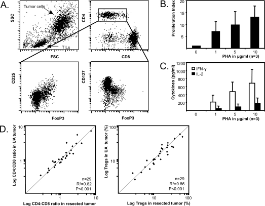
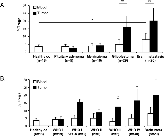
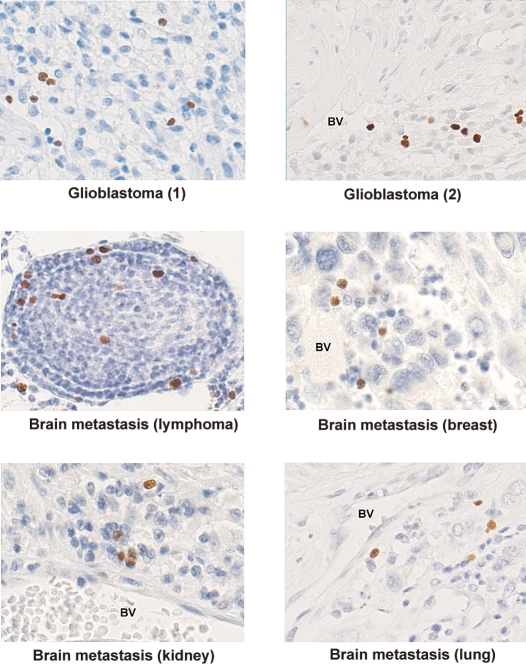
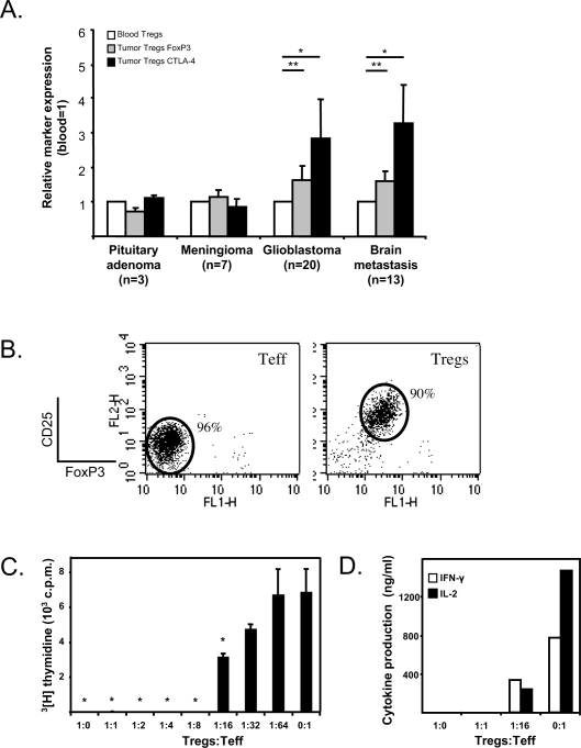
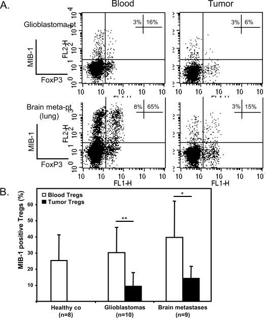
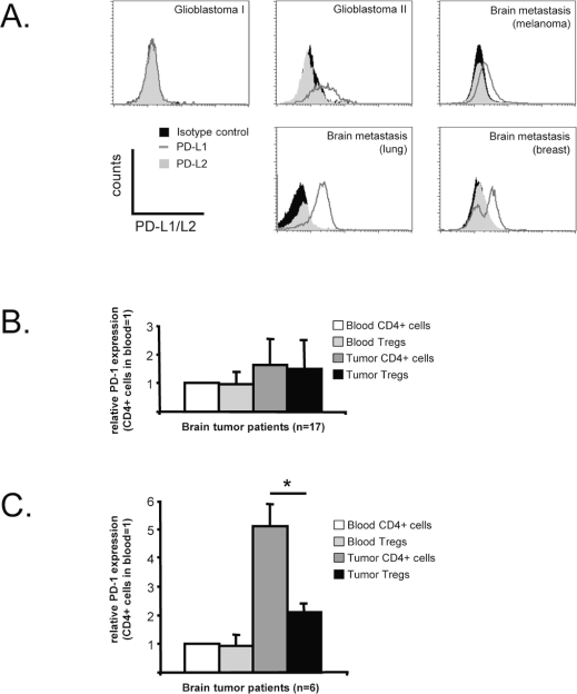
The brain is a specialized immune site representing a unique tumor microenvironment. The availability of fresh brain tumor material for ex vivo analysis is often limited because large parts of many brain tumors are resected using ultrasonic aspiration. We analyzed ultrasonic tumor aspirates as a biosource to study immune suppressive mechanisms in 83 human brain tumors. Lymphocyte infiltrates in brain tumor tissues and ultrasonic aspirates were comparable with respect to lymphocyte content and viability. Applying ultrasonic aspirates, we detected massive infiltration of CD4+FoxP3+CD25(high) CD127(low) regulatory T cells (Tregs) in glioblastomas (n = 29) and metastatic brain tumors (n = 20). No Treg accumulation was observed in benign tumors such as meningiomas (n = 10) and pituitary adenomas (n = 5). A significant Treg increase in blood was seen only in patients with metastatic brain tumors. Tregs in high-grade tumors exhibited an activated phenotype as indicated by decreased proliferation and elevated CTLA-4 and FoxP3 expression relative to blood Tregs. Functional analysis showed that the tumor-derived Tregs efficiently suppressed cytokine secretion and proliferation of autologous intratumoral lymphocytes. Most tumor-infiltrating Tregs were localized in close proximity to effector T cells, as visualized by immunohistochemistry. Furthermore, 61% of the malignant brain tumors expressed programmed death ligand-1 (PD-L1), while the inhibitory PD-1 receptor was expressed on CD4+ effector cells present in 26% of tumors. In conclusion, using ultrasonic tumor aspirates as a biosource we identified Tregs and the PD-L1/PD-1 pathway as immune suppressive mechanisms in malignant but not benign human brain tumors.


Figures

Fig. 1.
Fig. 1.
Flow cytometry of ultrasonic aspirated (UA) glioblastoma material (A). Lymphocytes can be differentiated into CD4+ cells (25%), CD8+ cells (65%), and a population mainly consisting of B cells (data not shown). CD4+ cells can further be characterized into CD4+FoxP3+ CD25highCD127low cells and CD4+FoxP3CD25lowCD127high cells. Glioblastoma brain tumor–infiltrating lymphocytes (TILs) proliferate (B) and produce IFN-γ and IL-2 (C) upon increasing phytohemagglutinin (PHA) stimulation (the average of 3 glioblastoma patients is shown). (D) CD4:CD8 ratio (left scatterplot) and percentage of Tregs (right scatterplot) in ultrasonic aspirated brain tumor and resected brain tumor are strongly correlated (n = 29 tumors).
Fig. 2.
Fig. 2.
Tregs accumulate in high-grade brain tumors. (A) Percentage of Tregs in blood and tumor of patients with brain tumors compared to healthy controls (co) (*p < 0.01, Student’s t-test, **p < 0.003, paired Student’s t-test). (B) Percentage of Tregs in brain tumors positively correlates with the WHO malignancy grade (*p < 0.01, Student’s t-test). WHO grade I subependymal giant-cell astrocytomas (SEGA) are represented as a separate group to illustrate the relatively high percentages of Tregs in this tumor. Abbreviation: co, control.
Fig. 3.
Fig. 3.
FoxP3 positive lymphocytes in paraffin-embedded brain tumors (236A/E7 antibody). Abbreviation: BV, blood vessel.
Fig. 4.
Fig. 4.
Tregs in high-grade brain tumors have an activated phenotype and strongly suppress proliferation of brain tumor–infiltrating lymphocytes (TILs). (A) FoxP3 and CTLA-4 expression are significantly increased in tumor-infiltrating Tregs compared to peripheral blood Tregs in patients with glioblastomas and patients with brain metastases (*p < 0.01; **p < 0.001, paired Student’s t-test). (B) Purified brain tumor–derived CD4+FoxP3CD25lowCD127high (effector T cells, or Teff) and CD4+FoxP3+CD25highCD127low (Tregs) were titrated in a suppression assay. (C) Proliferation was assessed by 3[H]thymidine incorporation (*p < 0.001, Student’s t-test) and (D) cytokines were analyzed in supernatants. One representative experiment out of three is shown. Abbreviation: c.p.m., count per minute.
Fig. 5.
Fig. 5.
Treg proliferation in vivo. (A) The percentages of MIB-1 (Ki-67 antibody)–positive Tregs (of total CD4+FoxP3+) and MIB-1– positive CD4+ cells (of total CD4+FoxP3) are indicated in the top right quadrants of the dot plots. Two representative patients are shown (Glioblastoma-pt and Brain meta-pt). (B) Overall MIB-1 expression on blood and intratumoral Tregs of healthy controls (co), patients with glioblastomas, and patients with brain metastases. The percentage of MIB-1–positive Tregs significantly decreases in the tumor (*p < 0.05; **p < 0.01, paired Student’s t-test). Abbreviations: glioblastoma-pt, patient with a glioblastoma; brain meta-pt, patient with a brain metastasis; co, control.
Fig. 6.
Fig. 6.
PD-L1/PD-1 expression in brain tumors. (A) PD-L1 and PD-L2 expression in five representative brain tumors. Sixty-one percent of the tumor samples expressed PD-L1 (dark gray line), which varied strongly between samples. PD-L2 expression (gray shaded histogram) was negative in all tested tumors (n = 23). (B) Blood lymphocytes do not express PD-1 on their cell surface. In 17 of 23 tested tumors, PD-1 expression in brain tumor–infiltrating lymphocytes (TILs) was not significantly upregulated. (C) In 6 of 23 tested tumors, PD-1 cell surface expression was specifically upregulated on tumor-infiltrating CD4+FoxP3 cells (*p = 0.005, paired Student’s t-test).

References

    1. Louis DN, Ohgaki H, Wiestler OD, Cavenee WK, editors. Tumours of the Central Nervous System. Lyon: IARC Press; 2007. - PMC - PubMed
    1. Legler JM, Ries LA, Smith MA, et al. Cancer surveillance series [corrected]: brain and other central nervous system cancers: recent trends in incidence and mortality. J Natl Cancer Inst. 1999;91:1382–1390. - PubMed
    1. Sawaya R, Ligon BL, Bindal RK. Management of metastatic brain tumors. Ann Surg Oncol. 1994;1:169–178. - PubMed
    1. Galea I, Bernardes-Silva M, Forse PA, van Rooijen N, Liblau RS, Perry VH. An antigen-specific pathway for CD8 T cells across the blood-brain barrier. J Exp Med. 2007;204:2023–2030. - PMC - PubMed
    1. Ransohoff RM, Kivisakk P, Kidd G. Three or more routes for leukocyte migration into the central nervous system. Nat Rev Immunol. 2003;3:569–581. - PubMed

Publication types