Review
. 2008 Dec;14(6):609-25.
doi: 10.1177/1073858408322675.

Presynaptic NMDA receptors: newly appreciated roles in cortical synaptic function and plasticity

Affiliations
Review

Presynaptic NMDA receptors: newly appreciated roles in cortical synaptic function and plasticity

Rebekah Corlew et al. Neuroscientist. 2008 Dec.

Abstract

Many aspects of synaptic development, plasticity, and neurotransmission are critically influenced by NMDA-type glutamate receptors (NMDARs). Moreover, dysfunction of NMDARs has been implicated in a broad array of neurological disorders, including schizophrenia, stroke, epilepsy, and neuropathic pain. Classically, NMDARs were thought to be exclusively postsynaptic. However, substantial evidence in the past 10 years demonstrates that NMDARs also exist presynaptically and that presynaptic NMDA receptors (preNMDARs) modulate synapse function and have critical roles in plasticity at many synapses. Here the authors review current knowledge of the role of preNMDARs in synaptic transmission and plasticity, focusing on the neocortex. They discuss the prevalence, function, and development of these receptors, and their potential modification by experience and in brain pathology.


Figures

Fig. 1
Fig. 1. PreNMDARs can enhance action potential-evoked neurotransmission
Evidence that preNMDARs modulate evoked transmitter release between pairs of L5 pyramidal neurons in primary visual cortex. (A) With postsynaptic NMDARs blocked by voltage-clamping the postsynaptic neuron at −90mV, preNMDAR blockade decreases EPSC amplitude and short-term synaptic depression. Sample recording showing averaged responses before (a) and during (b) application of APV. (B) APV wash-in reversibly decreased responses to 30 Hz spiking between pairs of L5 neurons recorded in voltage-clamp at −90 mV. Reproduced with permission from Sjöström and others (2003).
Fig. 2
Fig. 2. Synapse-specific expression of preNMDARs
PreNMDARs decrease synaptic strength and increase paired-pulse ratios at layer (L) 4-L2/3 synapses, but not at L2/3-L2/3 synapses or at L4-L4 synapses in the rodent somatosensory cortex. (A) Recording set-up for these experiments. Top: Representative experiment testing the effect of D-APV on AMPA-EPSCs evoked on the L2/3 cross-columnar pathway (L2/3-L2/3) and on L4-L2/3 inputs to the same postsynaptic cell. Amplitude of the first AMPA-EPSC on each pathway. Insets: Pairs of AMPA-EPSCs before (black) and during (grey) D-APV application. Bottom: Mean effect of D-APV on first AMPA-EPSC amplitude for L2/3-L2/3 inputs (triangles) and simultaneously measured L4-L2/3 inputs (circles). Bars represent population means (* = p < 0.05). (B) Differential interference contrast image of example synaptically coupled L4 excitatory cells, with regular-spiking pattern for these cells. Postsynaptic EPSCs elicited by a pair of presynaptic spikes before (black) and after (grey) 50µM D-APV application for the regular-spiking pair above. Lack of effect of D-APV on amplitude of the first uEPSC for one representative cell pair. Mean effect of D-APV application on first uEPSC amplitude. Reproduced with permission from Brasier and Feldman (2008).
Fig. 3
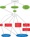
Fig. 3. Schematic of possible mechanisms for preNMDAR-enhancement of neurotransmitter release
There are three potential signals (green) emanating directly from the preNMDAR that could modulate release: direct Ca2+ entry, depolarization leading to the opening of VSCCs, and current-independent effects. The two ultimate mechanisms (blue) for expression of increased release are direct triggering of release by Ca2+ signals, or indirect signaling that changes the probability of release. Signaling to one of these ends could be carried out directly by Ca2+, or by several different intermediates (red): an amplified Ca2+ signal through calcium-induced calcium release (CICR), kinases/phosphatases that alter future Ca2+ entry, or increased Ca2+ -sensitivity of release machinery.
Fig. 4
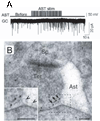
Fig. 4. Glutamate released from astrocytes can regulate neurotransmitter release
(A) Electrical stimulation (stim) of an astrocyte (AST) increases miniature excitatory synaptic currents recorded in a granule cell (GC) in the dentate gyrus of the hippocampus. (B) Electron micrographs showing NR2B gold particles in extrasynaptic membranes (arrows) of nerve terminals (Ter) making asymmetric synapses with dendritic spines (Sp) in the dentate molecular layer and an associated astrocytic process (Ast). Inset shows at higher magnification NR2B particles apposed to an astrocytic process that contains synaptic like microvesicles (arrowheads). Scale bars, 100 nm. Reproduced with permission from Jourdain and others (2007).
Fig. 5
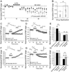
Fig. 5. The induction of some forms of long-term depression (LTD) requires preNMDARs
(A,B) Extracellularly evoked L4-L2/3 EPSPs in somatosensory cortex. (A) In L4-L2/3 somatosensory cortex connections, robust timing-dependent LTD can be induced in control experiments (closed circles) by repetitively pairing postsynaptic action potentials followed closely in time with an EPSP (pairing protocol is indicated by gray bar). The LTD is observed even when postsynaptic NMDARs are blocked with postsynaptic internal MK-801 (open circles), but not when APV is bath applied (not shown). (B) Bath-applying the endocannabinoid agonist AEA induces a lasting LTD at L4-L2/3 synapses (indicated by a reduction of EPSP slope, normalized to baseline). Prior blockade of pre- and post-synaptic NMDARs with bath applied D-APV blocks this AEA-induced LTD (no reduction in EPSP slope from baseline), while postsynaptic NMDAR blockade with MK-801 does not block AEA-induced LTD (not shown). (C–H) Synaptically coupled L4-L2/3 excitatory pairs in somatosensory cortex. (C) Postsynaptic MK-801 (grey) completely blocks induction of timing-dependent long-term potentiation (tLTP), while control (no MK-801) neurons (black) exhibit normal LTP. Inset, EPSP before (1) and 30 min after (2) the LTP pairing protocol. (D) Postsynaptic MK-801 did not block tLTD. Symbols and traces are presented as in C. (E) Summary of C, D. (F) During paired recordings, presynaptic MK-801 did not block the induction of tLTP. (G) Presynapitic MK-801 completely blocks tLTD. Symbols and traces are presented as in C. (H) Summary of F, G. Control tLTP refers to values obtained using extracellular stimulation. (E,H) The numbers of slices are shown in parentheses. All error bars are s.e.m. * = p < 0.05, ** = p < 0.01, Student’s t-test. (A,B) Reproduced with permission from Bender and others (2006), (C–H) reproduced with permission from Rodríguez-Moreno and Paulsen (2008).
Fig. 6
Fig. 6. Two models showing preNMDAR involvement in the coincidence detection of spike timing-dependent LTD
(A) It has been proposed in L5 of visual cortex (Sjöström and others 2003) that postsynaptic action potentials trigger the release of endocannabinoids in a mechanism similar to short-term depression caused during depolarization-induced suppression of inhibition. When presynaptic activity follows this cannabinoid release in a short time window, it coincidently activates preNMDARs and presynaptic CB1 receptors to trigger LTD induction. Reproduced with permission from Sjöström and others 2003. (B) Schematic for a possible postsynaptic coincidence detector for CB1-mediated tLTD. Black, pathway for mGluR-dependent cannabinoid synthesis. Red, pathway for VSCC- and calcium-dependent cannabinoid synthesis. Purple, potential synergistic pathways that increase cannabinoid production in response to appropriately timed pre- and postsynaptic spikes. Presynaptic NMDARs play a modulatory role in LTD in this hypothesis. Schematic based on data in Bender and others 2006.
Fig. 7
Fig. 7. Evidence for a developmental reduction in preNMDAR functions
(A) D,L-APV (or D-APV where indicated) strongly reduced AMPAR-mediated mEPSC frequency from baseline in L2/3, L4, and L5 pyramidal cells in the visual cortex of mice at P7–20 but not older mice. Sample sizes are given within the bars. (B) Immuno-electon microscopy for the obligatory NR1 subunit of the NMDAR reveals a developmental decrease in presynaptic, but not postsynaptic, NR1. Electron micrograph in L2/3 of visual cortex of a (B1) P16 mouse, demonstrating an NR1-positive terminal (t+) forming a synapse onto a NR1 positive spine (s+) and (B2) from a P27 mouse, demonstrating an unlabeled terminal (t-) forming a synapse onto a labeled dendrite (d+). Scale bar 250 nm. (C) Scatter plot from four mice (2 at each age) quantifying the selective loss of presynaptic, but not postsynaptic, NR1 over development. Reproduced with permission from Corlew and others (2007).
Fig. 8
Fig. 8. Developmental and activity-dependent regulation of preNMDAR functions
Presynaptic NR2B–containing receptors enhance spontaneous release in the entorhinal cortex of young rats and epileptic adults, but not in normal adults. (A–C) Voltage-clamp recordings of sEPSCs in a layer 5 neurons in a slice from (A) a 4-week-old rat, (B) a 5-month-old rat, and (C) and a 5-month-old epileptic rat. Blockade of NR2B receptors with Ro 25–6981 decreases the frequency of sEPSCs only in young and epileptic rats. Postsynaptic NMDARs are blocked by intracellular MK-801. (D–F) Corresponding pooled data for inter-event interval of the sEPSCs from control and Ro 25–6981 recordings. Reproduced with permission from Yang and others (2006).
Box 1
Box 1
Box 2
Box 2

References

    1. Allen CB, Celikel T, Feldman DE. Long-term depression induced by sensory deprivation during cortical map plasticity in vivo. Nat Neurosci. 2003;6:291–299. - PubMed
    1. Aoki C, Venkatesan C, Go CG, Mong JA, Dawson TM. Cellular and subcellular localization of NMDA-R1 subunit immunoreactivity in the visual cortex of adult and neonatal rats. J Neurosci. 1994;14:5202–5222. - PMC - PubMed
    1. Avanzini G, Franceschetti S. Cellular biology of epileptogenesis. Lancet Neurol. 2003;2:33–42. - PubMed
    1. Awatramani GB, Price GD, Trussell LO. Modulation of transmitter release by presynaptic resting potential and background calcium levels. Neuron. 2005;48:109–121. - PubMed
    1. Bardoni R, Torsney C, Tong CK, Prandini M, MacDermott AB. Presynaptic NMDA receptors modulate glutamate release from primary sensory neurons in rat spinal cord dorsal horn. J Neurosci. 2004;24:2774–2781. - PMC - PubMed

Publication types

MeSH terms

Substances

LinkOut - more resources