. 2009 Feb;77(2):632-41.
doi: 10.1128/IAI.01050-08. Epub 2008 Nov 24.

Vaccination with proteus toxic agglutinin, a hemolysin-independent cytotoxin in vivo, protects against Proteus mirabilis urinary tract infection

Affiliations

Vaccination with proteus toxic agglutinin, a hemolysin-independent cytotoxin in vivo, protects against Proteus mirabilis urinary tract infection

Praveen Alamuri et al. Infect Immun. 2009 Feb.

Abstract

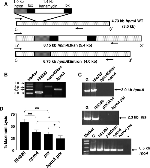
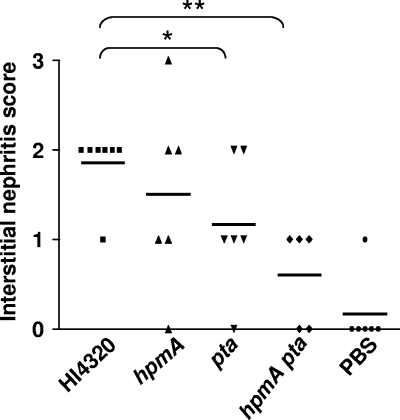
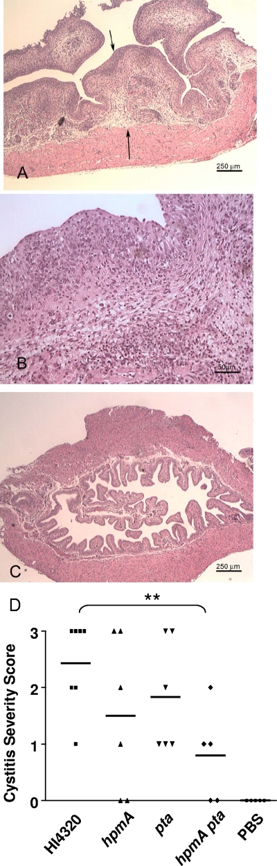
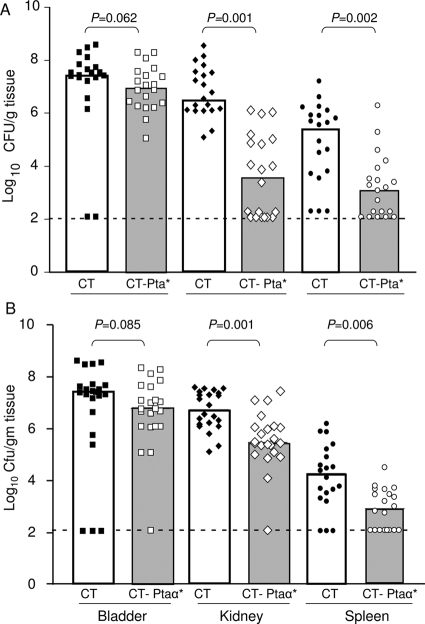
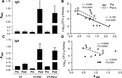
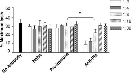
Complicated urinary tract infections (UTI) caused by Proteus mirabilis are associated with severe pathology in the bladder and kidney. To investigate the roles of two established cytotoxins, the HpmA hemolysin, a secreted cytotoxin, and proteus toxic agglutinin (Pta), a surface-associated cytotoxin, mutant analysis was used in conjunction with a mouse model of ascending UTI. Inactivation of pta, but not inactivation of hpmA, resulted in significant decreases in the bacterial loads of the mutant in kidneys (P < 0.01) and spleens (P < 0.05) compared to the bacterial loads of the wild type; the 50% infective dose (ID(50)) of an isogenic pta mutant or hpmA pta double mutant was 100-fold higher (5 x 10(8) CFU) than the ID(50) of parent strain HI4320 (5 x 10(6) CFU). Colonization by the parent strain caused severe cystitis and interstitial nephritis as determined by histopathological examination. Mice infected with the same bacterial load of the hpmA pta double mutant showed significantly reduced pathology (P < 0.01), suggesting that the additive effect of these two cytotoxins is critical during Proteus infection. Since Pta is surface associated and important for the persistence of P. mirabilis in the host, it was selected as a vaccine candidate. Mice intranasally vaccinated with a site-directed (indicated by an asterisk) (S366A) mutant purified intact toxin (Pta*) or the passenger domain Pta-alpha*, each independently conjugated with cholera toxin (CT), had significantly lower bacterial counts in their kidneys ( P = 0.001) and spleens (P = 0.002) than mice that received CT alone. The serum immunoglobulin G levels correlated with protection (P = 0.03). This is the first report describing the in vivo cytotoxicity and antigenicity of an autotransporter in P. mirabilis and its use in vaccine development.


Figures

FIG. 1.
FIG. 1.
Construction of hpmA and hpmA pta mutants and estimation of cytotoxicity. (A) Schematic diagram of the 4.73-kb hpmA gene, showing the site of inactivation by a kanamycin cassette. The approximate sizes of the hpmA gene, hpmAΩkan, and hpmAΩintron are indicated; the sizes of the corresponding PCR products amplified using the internal primers HpmAF and HpmAR (arrows) are indicated in parentheses. WT, wild type. (B) Insertion and excision of the kanamycin cassette in the hpmAΩkan and hpmAΩintron strains, respectively, was confirmed by amplification of hpmA by PCR. (C) Inactivation of hpmA in the hpmAΩkan and hpmAΩintron strains (top) and inactivation of pta in the hpmA pta double mutant (middle) were confirmed by RT-PCR using gene-specific primers. rpoA, encoding RNA polymerase A, was used as the expression control (bottom). The sizes of PCR products are indicated on the right. Lane G, HI4320 genomic DNA, lane −, no RT added to the cDNA from strain HI4320. (D) P. mirabilis HI4320 and the mutants were independently cultured until mid-exponential phase (optical density at 600 nm, 0.6 to 0.7) in alkaline LB (pH 8.5) supplemented with CaCl2 and 5% glycerol. A washed suspension of bacteria (106 CFU) in PBS was laid over a confluent monolayer of bladder cells and incubated for 4 h. The extent of bladder cell lysis (determined by the quantitative LDH release assay) is expressed as a percentage of the maximum cell lysis obtained by Triton X-100 treatment. The data are the means and standard errors of three independent experiments, each conducted in triplicate. *, P < 0.05; **, P < 0.01.
FIG. 2.
FIG. 2.
Mice were independently transurethrally inoculated with P. mirabilis HI4320 or the mutant constructs. After 7 days of infection, a kidney from each mouse was fixed, and the sections were stained for histopathological evaluation. (A) Kidney from a mouse inoculated with the hpmA mutant of P. mirabilis. Multiple foci of inflammation and necrosis are scattered throughout the cortex (arrows). The arrowheads delineate the border between more-normal tissue (left) and necrotic tissue (right). (Inset) Overview of the entire kidney, indicating the location of the section. (B) Higher magnification of panel A showing a focus of necrosis, neutrophilic inflammation, and bacterial colonies. (C) Normal kidney from an uninfected mouse. (D) Higher magnification of panel C. (E) Kidney from a mouse inoculated with wild-type P. mirabilis HI4320. A mild lymphocytic infiltrate is present adjacent to the renal pelvis (arrow). (F) Higher magnification of panel E showing lymphocytic infiltrate.
FIG. 3.
FIG. 3.
Interstitial nephritis caused by P. mirabilis HI4320 and isogenic mutants of this strain. Histopathology was scored as described in Materials and Methods. The horizontal bars indicate the means. P values were calculated by the one-tailed Mann-Whitney U test. *, P < 0.05; **, P < 0.01.
FIG. 4.
FIG. 4.
Histopathological evaluation of bladders from mice. (A) The urinary bladder obtained from a mouse after 7 days of infection with 5 × 10⁶ CFU of parent strain HI4320 was processed for histopathological evaluation as described in the text. The mucosa is thickened with edema and inflammatory cells (arrows). (B) Higher magnification of panel A showing neutrophils within the epithelium. (C) Bladder from a mouse that received PBS as a control. (D) Cystitis severity after inoculation of various P. mirabilis constructs. The horizontal bars indicate the means. P values were calculated by the one-tailed Mann-Whitney U test. **, P < 0.01.
FIG. 5.
FIG. 5.
Immune protection from P. mirabilis challenge by vaccination with Pta* and Pta-α*. Mice were vaccinated and boosted with CT-Pta* (A) or CT-Pta-α* (B) as described in the text and were challenged intraurethrally on day 21 with 5 × 10⁷ CFU P. mirabilis HI4320. The numbers of P. mirabilis CFU in bladders, kidneys, and spleens of naïve mice (which received CT alone) and mice immunized with CT-Pta-α* or CT-Pta* were determined 1 week after bacterial challenge. Each symbol indicates the log10 CFU/g of tissue from an individual mouse. Samples in which colonization was undetectable were given a value of 2.1 log10 CFU/g of tissue (the limit of detection). The bars indicate medians. P values were determined using the one-tailed Mann-Whitney U test; a P value of <0.05 was considered significant.
FIG. 6.
FIG. 6.
Correlation of IgG and IgA titers with protection. The antibody responses in mice to intranasal vaccination with CT-Pta*, with CT-Pta-α*, or with CT alone were determined by an indirect ELISA. Serum IgG and urine IgA titers in pre- and postimmune sera and urine collected from each group of immunized and naïve mice were determined using a 1:512 dilution of serum or urine. Goat anti-rabbit IgA or IgG conjugated with alkaline phosphatase was used as the secondary antibody. The alkaline phosphatase activity was determined at A405 60 min after addition of the substrate. The means and standard errors for serum IgG (A) and urine IgA (C) titers for eight mice (with each experiment performed in duplicate) are indicated. The bacterial load recovered from immunized mice 1 week after challenge was tested to determine correlations with Pta*- or Pta-α*-specific serum IgG (B) and urine IgA (D) responses. Both the anti-Pta and anti-Pta-α serum IgG titers were inversely correlated with the bacterial load, whereas the inverse correlation was significant only for Pta-α*-specific urine IgA and was not significant for Pta*. P values were calculated for correlation coefficients, and a P value of <0.05 was considered significant. Spearman's rank correlation coefficients (r) are indicated. The diagonal lines indicate linear regression.
FIG. 7.
FIG. 7.
Neutralization of Pta by anti-Pta sera. LDH release was used as a measure of bladder cell lysis after inoculation of P. mirabilis preincubated for 3 h with different dilutions of sera. The y axis indicates bladder cell lysis expressed as a percentage of the maximum lysis (after treatment with 2% [wt/vol] Triton X-100) normalized to the bladder cell lysis by the pta mutant. No antibody, strain HI4320 incubated with no serum; Naive, strain HI430 preincubated with sera collected from CT-immunized mice on day 21; Pre-immune, strain HI430 preincubated with sera collected from mice on day 1 prior to vaccination with Pta*; Anti-Pta, strain HI430 preincubated with sera collected on day 21 from mice immunized with Pta*. The one-tailed Mann-Whitney U test was used to determine P values. *, P < 0.05.
FIG. 8.
FIG. 8.
Prevalence of Pta in P. mirabilis isolates. Eight catheter-associated strains (C1 to C8) and eight fecal isolates (F1 to F8) were independently cultured to mid-exponential phase in LB (pH 8.5) supplemented with 10 mM CaCl2 and 5% glycerol. (A) Expression of pta in catheter-associated strains was determined by RT-PCR using pta-specific primers; cDNA obtained from each strain was used as a template. (B) Prevalence of pta in the genomes of fecal isolates as determined by PCR of genomic DNA. (C) Expression of pta in the strains as determined by RT-PCR using cDNA obtained from each isolate and pta-specific primers. rpoA, encoding RNA polymerase A, was used as an expression control. cDNA from strain HI4320 was used as the reference. Lane G, genomic DNA from strain HI4320 used as a template; lanes −, no RT added; lanes +, RT added. Sizes (in kb) are indicated on the right.

References

    1. Alamuri, P., and H. L. Mobley. 2008. A novel autotransporter of uropathogenic Proteus mirabilis is both a cytotoxin and an agglutinin. Mol. Microbiol. 68997-1017. - PubMed
    1. Allison, C., H. C. Lai, and C. Hughes. 1992. Co-ordinate expression of virulence genes during swarm-cell differentiation and population migration of Proteus mirabilis. Mol. Microbiol. 61583-1591. - PubMed
    1. Bahrani, F. K., D. E. Johnson, D. Robbins, and H. L. Mobley. 1991. Proteus mirabilis flagella and MR/P fimbriae: isolation, purification, N-terminal analysis, and serum antibody response following experimental urinary tract infection. Infect. Immun. 593574-3580. - PMC - PubMed
    1. Braun, V., and T. Focareta. 1991. Pore-forming bacterial protein hemolysins (cytolysins). Crit. Rev. Microbiol. 18115-158. - PubMed
    1. Cover, T. L., and S. R. Blanke. 2005. Helicobacter pylori VacA, a paradigm for toxin multifunctionality. Nat. Rev. Microbiol. 3320-332. - PubMed

Publication types