. 2008 Dec 1;183(5):923-32.
doi: 10.1083/jcb.200808050. Epub 2008 Nov 24.

Molecular architecture of inner dynein arms in situ in Chlamydomonas reinhardtii flagella

Affiliations

Molecular architecture of inner dynein arms in situ in Chlamydomonas reinhardtii flagella

Khanh Huy Bui et al. J Cell Biol. .

Abstract

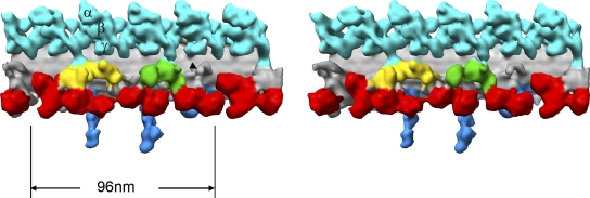
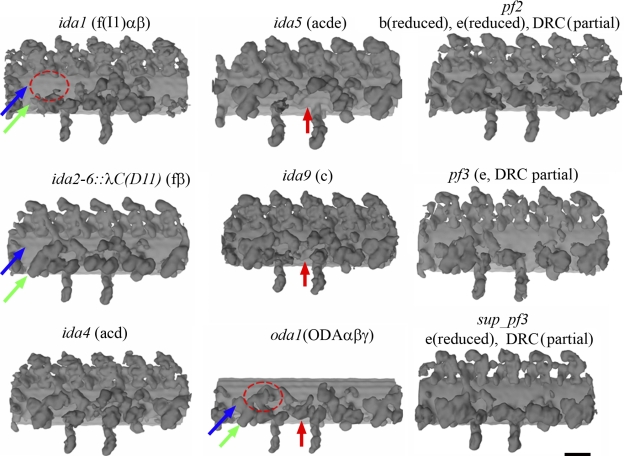
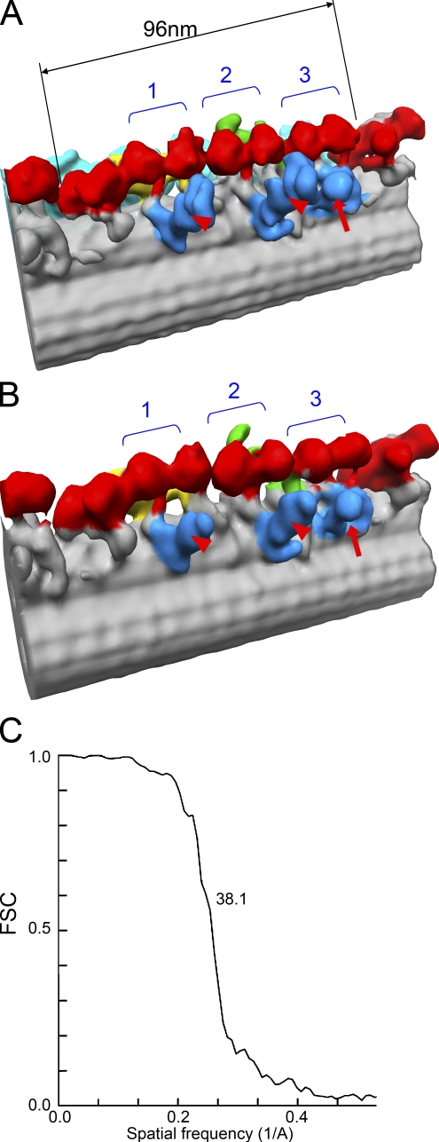
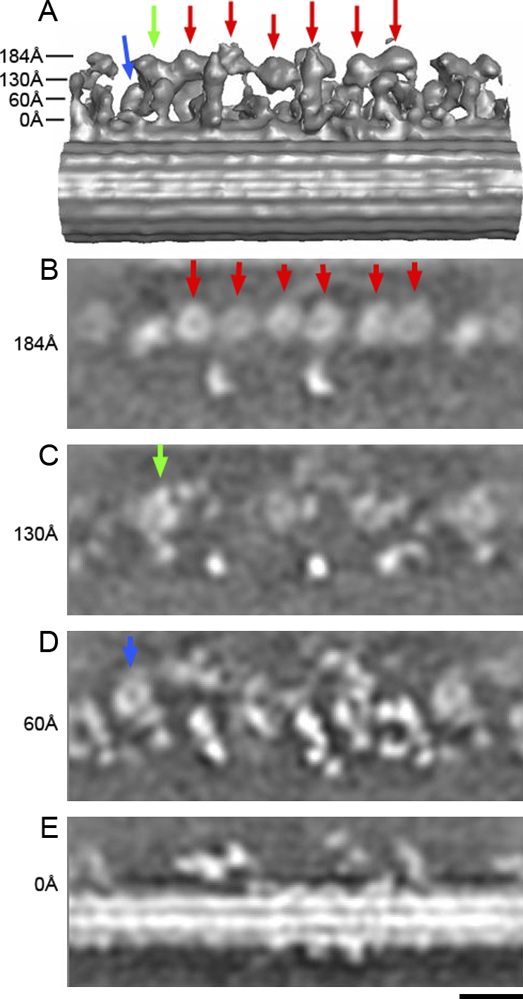
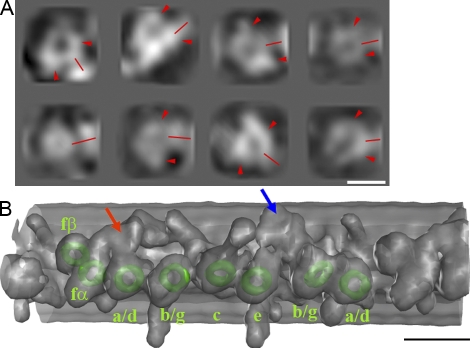
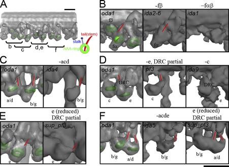
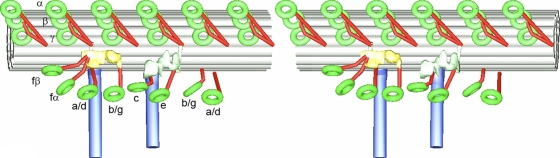
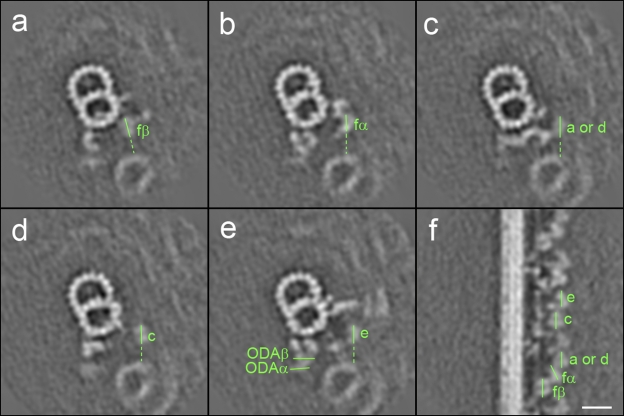
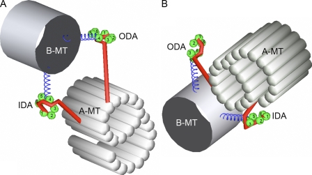
The inner dynein arm regulates axonemal bending motion in eukaryotes. We used cryo-electron tomography to reconstruct the three-dimensional structure of inner dynein arms from Chlamydomonas reinhardtii. All the eight different heavy chains were identified in one 96-nm periodic repeat, as expected from previous biochemical studies. Based on mutants, we identified the positions of the AAA rings and the N-terminal tails of all the eight heavy chains. The dynein f dimer is located close to the surface of the A-microtubule, whereas the other six heavy chain rings are roughly colinear at a larger distance to form three dyads. Each dyad consists of two heavy chains and has a corresponding radial spoke or a similar feature. In each of the six heavy chains (dynein a, b, c, d, e, and g), the N-terminal tail extends from the distal side of the ring. To interact with the B-microtubule through stalks, the inner-arm dyneins must have either different handedness or, more probably, the opposite orientation of the AAA rings compared with the outer-arm dyneins.


Figures

Figure 1.
Figure 1.
Surface rendering (stereo view) representation of the three-dimensional structure of the C. reinhardtii flagellum. The representation includes the A-microtubule, outer dynein arms (light blue), inner dynein arms (red), the IC–LC of the inner dynein arms (yellow), the DRC (green), and radial spokes (RS; blue) colored based on Porter (1996) within a 96-nm repeat. AAA rings of the three heavy chains in one outer dynein arm are also indicated. The black arrowhead indicates the candidate of nexin.
Figure 2.
Figure 2.
Three-dimensional structure of mutants. Components missing in each mutant are indicated in parenthesis. In ida1, ida2-6∷λC(D11), ida5, ida9, and oda1 mutants, the locations of a dynein f dimer (blue and green) and dynein c (red) are indicated by arrows, whereas the location of the LC–IC complex of dynein f is encircled by red dotted circles. Bar, 20 nm.
Figure 3.
Figure 3.
Three-dimensional reconstruction of flagella from the wild type and a mutant. Surface rendering representations of the wild type (A) and oda1 (B) flagellum seen from a different angle compared with Fig. 1. Six of eight dynein heavy chains form three dyads in the inner dynein arm. (left) The proximal end. (right) The distal end. Two radial spokes (red arrowheads) and one protrusion (red arrows) are associated with the three dyads, respectively. Stereo pairs are shown in Fig. S1 (available at http://www.jcb.org/cgi/content/full/jcb.200808050/DC1). Online supplemental videos are also available. (C) Fourier shell correlation curve of the oda1 mutant.
Figure 4.
Figure 4.
Horizontal sections from the three-dimensional density map of the oda1 mutant at various heights. Arrows in A indicate the heights in the surface rendering model. The six AAA rings (to be assigned as dynein a, b, c, d, e, and g later), and dynein fα and fβ are shown by red, green, and blue arrows, respectively. In horizontal sections (B, C, and D), six of the eight DHC rings are at the same height, whereas two are located closer to the A-microtubule. Bar, 20 nm.
Figure 5.
Figure 5.
Detection of the AAA rings in the density map of the oda1 mutant. (A) The eight rings detected in the 96-nm repeat of the oda1 mutant as projections. (left) Proximal end. (right) Distal end. N-terminal tails, which project out between two bulky densities (arrowheads), are shown by red lines. (B) Orientations of the eight rings are shown. Isoforms of dynein heavy chains are indicated by green rings based on Fig. 2. LC–IC complex and DRC are shown by red and blue arrows, respectively. A stereo view of B is shown in Fig. S1 (available at http://www.jcb.org/cgi/content/full/jcb.200808050/DC1). Bars: (A) 10 nm; (B) 20 nm.
Figure 6.
Figure 6.
Detailed comparisons between oda1 and other mutants reveal the location of the AAA rings and the N-terminal tails of the eight dynein heavy chains. In each panel, the orientation is slightly different in order to show the rings and the tails, but the distal end is always at the right (the proximal side is at the left). Approximate positions and the model of one dynein heavy chain (right bottom; green, AAA ring; red, N-terminal tail [stem]; blue, coiled-coil stalk and microtubule-binding domain) are shown in A. The rings and the tails are shown by green rings and red lines, respectively. Two tails of the dynein f dimer merge (B), whereas pairs of heavy chains belonging to one dyad have a colinear architecture, with the N-terminal tails projecting from the distal sides of the rings without merging (C–F). Bar, 20 nm.
Figure 7.
Figure 7.
Schematic diagram of the arrangement of the inner and outer dynein arms (stereo view). The rings belonging to the inner and outer dynein arms are shown as green rings. The N-terminal tails are shown in red.
Figure 8.
Figure 8.
Sections of the density map of the wild type. (a–e) Vertical sections (perpendicular to the microtubule) of the wild-type flagellum. AAA rings of the heavy chains of inner and outer dynein arms are marked by green lines. The solid lines show the sections of the planes of the rings. Corresponding locations and the orientations of the inner arm dynein rings are shown in the longitudinal section (f; so the lines in a–e and the lines in f are perpendicular to each other). (a) The section including dynein fβ. (b) Dynein fα. The distance from a is 75 Å. (c) Dynein a or d (180 Å from a). (d) Dynein c (450 Å from a). (e) Dynein e (580 Å). Dotted lines are extensions in the same plane as the rings and are presumed to represent the orientation of the stalks. ODA, outer dynein arm. Bar, 20 nm.
Figure 9.
Figure 9.
Schematic diagram of the conformation of heavy chains in the inner and outer dynein arms. (A) The view from the B-microtubule (B-MT). (left) Proximal side. (right) Distal side. (B) The view from the A-microtubule (A-MT). (left) Distal side. (right) Proximal side. The AAA domains are marked. The assignment and the conformation of the six AAA domains (green balls) in each ring, the coiled-coil stalks (blue helices), and the N-terminal tails (red rods) are based on Burgess et al. (2004). The likely arrangement of the six AAA domains is indicated based on the two-dimensional averaging of dynein c (Burgess, S., personal communication). Combining this indexing and three-dimensional reconstruction of cytoplasmic dynein (Samso and Koonce, 2004), we speculate that the tail wraps around the AAA ring, as shown here. IDA, inner dynein arm; ODA, outer dynein arm.

References

    1. Avolio, J., A.N. Glazzard, M.E. Holwill, and P. Satir. 1986. Structures attached to doublet microtubules of cilia: computer modeling of thin-section and negative-stain stereo images. Proc. Natl. Acad. Sci. USA. 83:4804–4808. - PMC - PubMed
    1. Bozkurt, H.H., and D.M. Woolley. 1993. Morphology of nexin links in relation to interdoublet sliding in the sperm flagellum. Cell Motil. Cytoskeleton. 24:109–118. - PubMed
    1. Burgess, S.A., D.A. Carter, S.D. Dover, and D.M. Woolley. 1991. The inner dynein arm complex: compatible images from freeze-etch and thin section methods of microscopy. J. Cell Sci. 100:319–328. - PubMed
    1. Burgess, S.A., M.L. Walker, H. Sakakibara, P.J. Knight, and K. Oiwa. 2003. Dynein structure and power stroke. Nature. 421:715–718. - PubMed
    1. Burgess, S.A., M.L. Walker, H. Sakakibara, H. Oiwa, and P.J. Knight. 2004. The structure of dynein-c by negative stain electron microscopy. J. Struct. Biol. 146:205–216. - PubMed

Publication types