. 2009 Apr;57(4):289-300.
doi: 10.1369/jhc.2008.952044. Epub 2008 Nov 24.

Assessment of apoptosis by immunohistochemistry to active caspase-3, active caspase-7, or cleaved PARP in monolayer cells and spheroid and subcutaneous xenografts of human carcinoma

Affiliations

Assessment of apoptosis by immunohistochemistry to active caspase-3, active caspase-7, or cleaved PARP in monolayer cells and spheroid and subcutaneous xenografts of human carcinoma

Aude Bressenot et al. J Histochem Cytochem. 2009 Apr.

Abstract

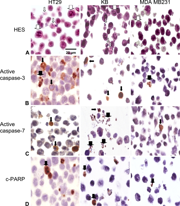
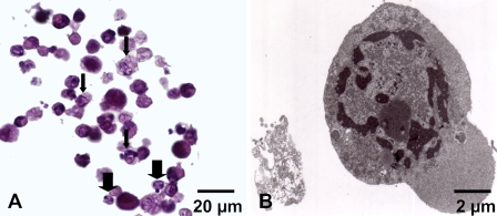
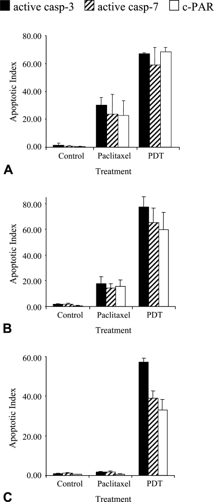
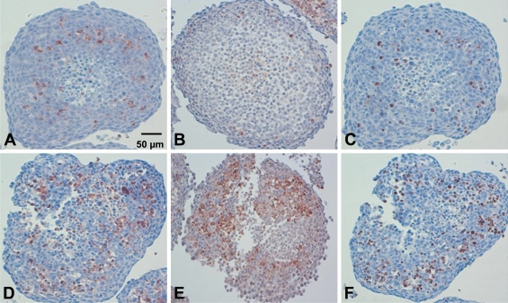
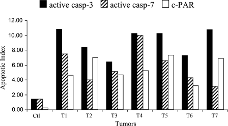
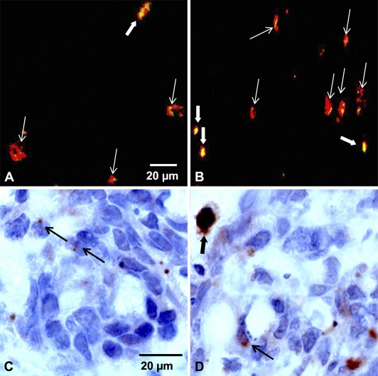
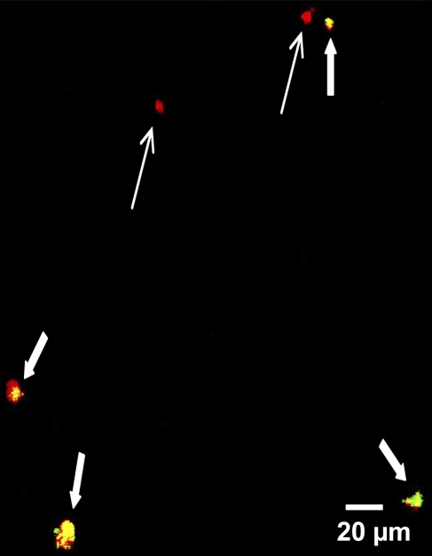
Immunohistochemistry to active caspase-3, recently recommended for apoptosis detection, is inappropriate to detect apoptosis involving caspase-7. Cleavage of poly-ADP-ribose polymerase 1 (PARP-1), a major substrate of both caspases, is a valuable marker of apoptosis. Apoptosis evaluation induced in vitro either by paclitaxel or by photodynamic treatment (PDT) with Foscan in HT29 or KB monolayer cells and HT29 spheroids yielded a close percentage of labeled cells whatever the antibody used, whereas in control specimens, cleaved PARP (c-PARP) immunostaining failed to detect apoptosis as efficiently as active caspase-3 or -7 immunostaining. Studies in MDA-MB231 monolayer cells and HT29 xenografts either subjected or not subjected to Foscan-PDT resulted in a significant higher number of active caspase-3-labeled cells, although immunofluorescence analysis showed c-PARP and active caspase-3 perfectly colocalized in tumors. A restricted expression of c-PARP was obvious in the greater part of caspase-3 expressing cells from control tumor, whereas photosensitized tumors showed a higher number of cells expressing large fluorescent spots from both active caspase-3 and c-PARP. These results support the assumption that c-PARP expression was dependent on treatment-induced apoptosis. The absence of caspase-7 activation in some caspase-3-expressing cells undergoing Foscan-PDT shows the relevance of using antibodies that can discriminate caspase-dependent apoptotic pathways.


Figures

Figure 1
Figure 1
Apoptosis and apoptosis-related protein detection in HT29, KB, and MDA-MB231 monolayer cells subjected to 0.1 μM paclitaxel for 48 hr. (A) Paraffin sections were stained with standard coloration hematoxylin-eosin-safran. Note specific morphological features of apoptosis in HT29 and KB monolayers cells (thin white arrows). Numerous mitotic cells were observed in the three cell lines (thick white arrows). (B) Active caspase-3 labeling. Note signal cytoplasmic location (thin black arrows) and focally nuclear signal translocation (thick black arrows). (C) Active caspase-7 labeling. Cytoplasm location (thin black arrows) and apoptotic bodies (thick black arrows) were observed. (D) Cleaved poly-ADP-ribose polymerase (c-PARP) labeling. Nuclear location was noticed (thick black arrows). Harris' hematoxylin counterstain.
Figure 2
Figure 2
Apoptosis in MDA-MB231 monolayer cells subjected to Foscan-photodynamic treatment (PDT). (A) Paraffin sections were stained with standard coloration HES. Note specific morphological features of apoptosis such as condensed eosinophilic cytoplasm and clumped marginated chromatin (thin arrows) or apoptotic bodies (thick arrows). (B) Transmission electron microscopy. Ultrastructural study showed an MDA-MB231 cell with a nuclear marginated chromatin and an increase in the cytosol density typical of apoptosis.
Figure 3
Figure 3
Apoptotic index from active caspase-3, active caspase-7, or c-PARP staining applied on control or treated HT29 (A), KB (B), and MDA-MB231 (C) cells. Cells were treated either by paclitaxel or Foscan-PDT. Results are expressed as mean apoptotic index (AI) ± SD of three experiments determined from at least 2000 tumor cells in each section. *Significantly different values (p<0.05).
Figure 4
Figure 4
IHC on paraffin sections of HT29 spheroids to active caspase-3 (A,D), active caspase-7 (B,E), or c-PARP (C,F) applied without treatment (A–C) or subjected to Foscan-PDT (D–F). Note the distribution of apoptotic cells at the periphery of necrotic area. Loss of cell cohesion was noticeable in spheroids subjected to Foscan-PDT (D–F). Harris' hematoxylin counterstain.
Figure 5
Figure 5
Apoptotic index from active caspase-3, active caspase-7, or c-PARP staining applied on HT29 subcutaneous xenografts subjected to Foscan (0.3 mg/kg, IV) photosensitization (10 J/cm2, 30 mW/cm2, 24 hr after injection). Note the variability between tumors. AIs were determined from at least 2000 tumor cells in each section.
Figure 6
Figure 6
Merging of immunofluorescence images obtained from control tumors (A) or Foscan (0.3 mg/kg, IV)-photosensitized (10 J/cm2, 30 mW/cm2) HT29 subcutaneous xenografts (B) frozen 24 hr after treatment. Frozen sections were first subjected to active caspase-3 (red fluorescence) and then to c-PARP (green fluorescence). Colocalization between both markers showed large yellow spots (thick white arrows) or tiny yellow spots (thin white arrows) caused by c-PARP expression. c-PARP IHC on paraffin sections of control tumors (C) and photosensitized tumors (D). Thin black arrows indicate limited expression of c-PARP, whereas the thick black arrow indicates a large expression of c-PARP in nuclei of apoptotic cells.
Figure 7
Figure 7
Merging of immunofluorescence images obtained from Foscan (0.3 mg/kg, IV)-photosensitized (10 J/cm2, 30 mW/cm2) HT29 subcutaneous xenografts frozen 24 hr after treatment. Frozen sections were first subjected to active caspase-3 (red fluorescence) and then to active caspase-7 (green fluorescence). Colocalization between both markers showed yellow spots (thick arrows). Red spots (thin arrows) indicate active caspase-3 expression only.

References

    1. Alvarez-Gonzalez R, Spring H, Muller M, Burkle A (1999) Selective loss of poly(ADP-ribose) and the 85-kDa fragment of poly(ADP-ribose) polymerase in nucleoli during alkylation-induced apoptosis of HeLa cells. J Biol Chem 274:32122–32126 - PubMed
    1. Coutier S, Bezdetnaya LN, Foster TH, Parache RM, Guillemin F (2002) Effect of irradiation fluence rate on the efficacy of photodynamic therapy and tumor oxygenation in meta-tetra (hydroxyphenyl) chlorin (mTHPC)-sensitized HT29 xenografts in nude mice. Radiat Res 158:339–345 - PubMed
    1. D'Amours D, Sallmann FR, Dixit VM, Poirier GG (2001) Gain-of-function of poly(ADP-ribose) polymerase-1 upon cleavage by apoptotic proteases: implications for apoptosis. J Cell Sci 114:3771–3778 - PubMed
    1. Davidson DJ, Haskell C, Majest S, Kherzai A, Egan DA, Walter KA, Schneider A, et al. (2005) Kringle 5 of human plasminogen induces apoptosis of endothelial and tumor cells through surface-expressed glucose-regulated protein 78. Cancer Res 65:4663–4672 - PubMed
    1. Decker P, Muller S (2002) Modulating poly (ADP-ribose) polymerase activity: potential for the prevention and therapy of pathogenic situations involving DNA damage and oxidative stress. Curr Pharm Biotechnol 3:275–283 - PubMed

MeSH terms