Review
. 2008 Dec 1;112(12):4384-99.
doi: 10.1182/blood-2008-07-077982.

Classification of lymphoid neoplasms: the microscope as a tool for disease discovery

Affiliations
Review

Classification of lymphoid neoplasms: the microscope as a tool for disease discovery

Elaine S Jaffe et al. Blood. .

Abstract

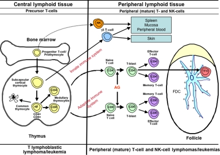
In the past 50 years, we have witnessed explosive growth in the understanding of normal and neoplastic lymphoid cells. B-cell, T-cell, and natural killer (NK)-cell neoplasms in many respects recapitulate normal stages of lymphoid cell differentiation and function, so that they can be to some extent classified according to the corresponding normal stage. Likewise, the molecular mechanisms involved the pathogenesis of lymphomas and lymphoid leukemias are often based on the physiology of the lymphoid cells, capitalizing on deregulated normal physiology by harnessing the promoters of genes essential for lymphocyte function. The clinical manifestations of lymphomas likewise reflect the normal function of lymphoid cells in vivo. The multiparameter approach to classification adopted by the World Health Organization (WHO) classification has been validated in international studies as being highly reproducible, and enhancing the interpretation of clinical and translational studies. In addition, accurate and precise classification of disease entities facilitates the discovery of the molecular basis of lymphoid neoplasms in the basic science laboratory.


Figures

Figure 1
Figure 1
Schematic diagram illustrating evolution of the entity MCL. MCL was recognized in Kiel classification and modified Rappaport classification as centrocytic lymphoma and lymphocytic lymphoma of intermediate differentiation (IDL), respectively. Precise criteria for the distinction from other morphologically similar lymphomas were lacking. The recognition of a characteristic immunophenotype (CD5+, CD23, CD10, monoclonal B cell) helped better define the entity. The identification of the t(11;14) resulting in CCNDI/IGH translocation in virtually all cases of MCL led to the use of cyclin D1 detection by immunohistochemistry for diagnosis. In addition, secondary genetic events such as p53 and p16 deletion/mutation were identified in high-grade variants of MCL, which had been recognized histologically as the “blastoid” subtype. The data derived from immunophenotypic and genetic studies are integrated, culminating in our current definition of the disease. PDL indicates poorly differentiated lymphocytic; WDL, well-differentiated lymphocytic; DHL, diffuse histiocytic lymphoma; and MZL, marginal zone lymphoma.
Figure 2
Figure 2
Diagrammatic representation of B-cell differentiation and relationship to major B-cell neoplasms. B-cell neoplasms correspond to stages of B-cell maturation, even though the precise cell counterparts are not known in all instances. Precursor B cells that mature in the bone marrow may undergo apoptosis or develop into mature naive B cells that, following exposure to antigen and blast transformation, may develop into short-lived plasma cells or enter the germinal center (GC), where somatic hypermutation and heavy chain class-switching occur. Centroblasts, the transformed cells of the GC, either undergo apoptosis or develop into centrocytes. Post-GC cells include both long-lived plasma cells and memory/marginal zone B cells. Most B cells are activated within the GC, but T cell–independent activation can take place outside of the GC and also probably leads to memory-type B cells. Monocytoid B cells, many of which lack somatic hypermutation, are not illustrated. AG indicates antigen; and FDC, folllicular dendritic cell. Red bar represents immunoglobulin heavy chain gene (IGH@) rearrangement; blue bar, immunoglobulin light chain gene (IGL) rearrangement; and black insertions in the red and blue bars indicate somatic hypermutation.
Figure 3
Figure 3
Diagrammatic representation of T-cell differentiation and function. Lymphoid progenitors enter the thymus where precursor T cells develop into varied types of naive T cells. The cells of the innate immune system include NK cells, γδ T cells, and NK-like T cells. These cells constitute a primitive type of immune response that lacks both specificity and memory. In the adaptive immune system, αβ T cells leave the thymus, where, upon exposure to antigen, they may undergo blast transformation and develop further into CD4+ and CD8+ effector and memory T cells. T cells of the adaptive immune system are heterogeneous and functionally complex, and include naive, effector (regulatory and cytotoxic), and memory T cells. Another specific type of effector T cells is the follicular helper T-cell that is found in GCs (TFH). Upon antigenic stimulation, T-cell responses may occur independent of the GC, or in the context of a GC reaction. The lymphomas of the innate immune system are predominantly extranodal in presentation, mirroring the distribution of the functional components of this system. T-cell lymphomas of the adaptive immune system present primarily in adults, and are mainly nodal in origin.
None
Elaine S. Jaffe
None
Nancy Lee Harris
None
Harald Stein
None
Peter G. Isaacson

References

    1. Swerdlow SH, Campo E, Harris NL, et al. WHO Classification of Tumours of Haematopoietic and Lymphoid Tissues. 4th ed. Lyon, France: International Agency for Research on Cancer; 2008.
    1. Kyle RA, Rajkumar SV. Multiple myeloma. Blood. 2008;111:2962–2972. - PMC - PubMed
    1. Swerdlow S, Habeshaw J, Murray L, Dhaliwal H, Lister T, Stansfeld A. Centrocytic lymphoma: a distinct clinicopathologic and immunologic entity. Am J Pathol. 1983;113:181. - PMC - PubMed
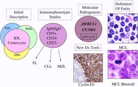
    1. Jaffe ES, Bookman MA, Longo DL. Lymphocytic lymphoma of intermediate differentiation–mantle zone lymphoma: a distinct subtype of B-cell lymphoma. Hum Pathol. 1987;18:877–880. - PubMed
    1. Stein H, Mason DY, Gerdes J, et al. The expression of the Hodgkin's disease associated antigen Ki-1 in reactive and neoplastic lymphoid tissue: evidence that Reed-Sternberg cells and histiocytic malignancies are derived from activated lymphoid cells. Blood. 1985;66:848–858. - PubMed

MeSH terms