. 2008 Nov 24;5(4):046005.
doi: 10.1088/1478-3975/5/4/046005.

The ribosome structure controls and directs mRNA entry, translocation and exit dynamics

Affiliations

The ribosome structure controls and directs mRNA entry, translocation and exit dynamics

Ozge Kurkcuoglu et al. Phys Biol. .

Abstract

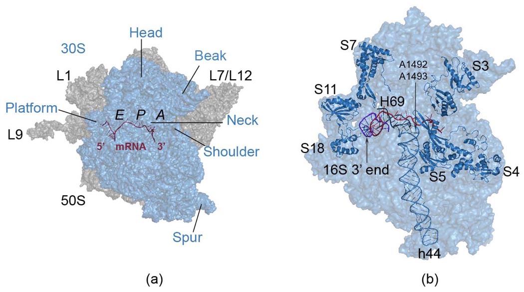
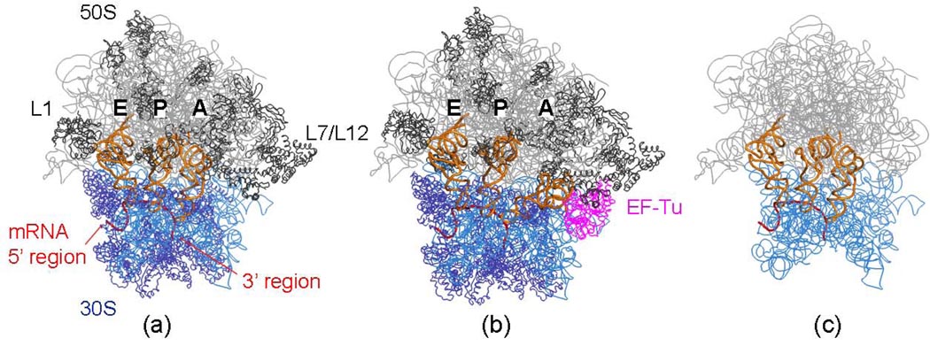
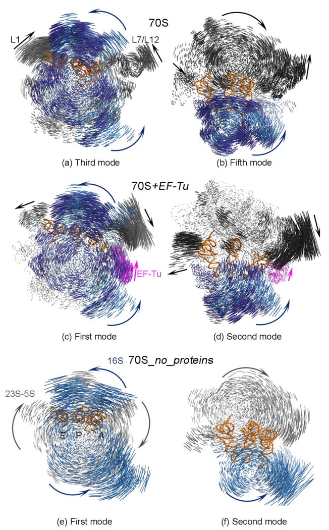
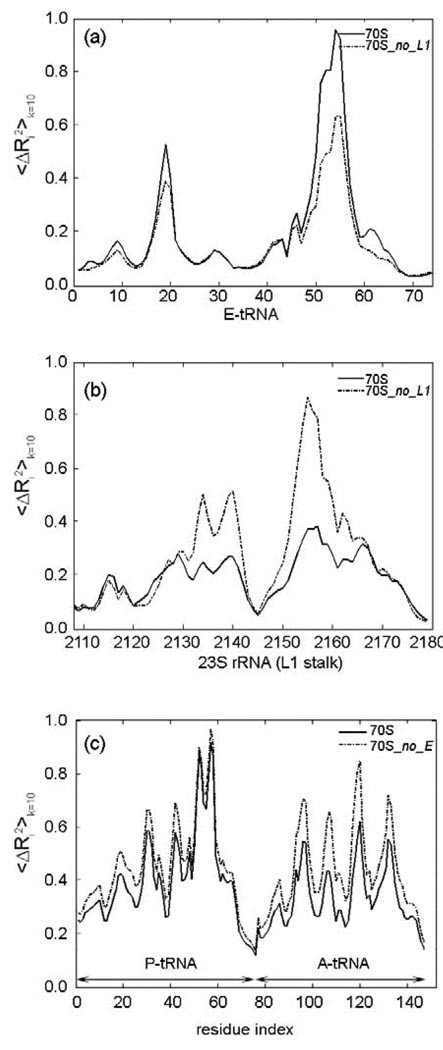
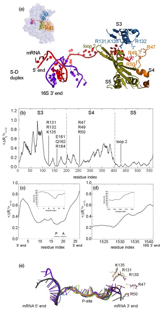
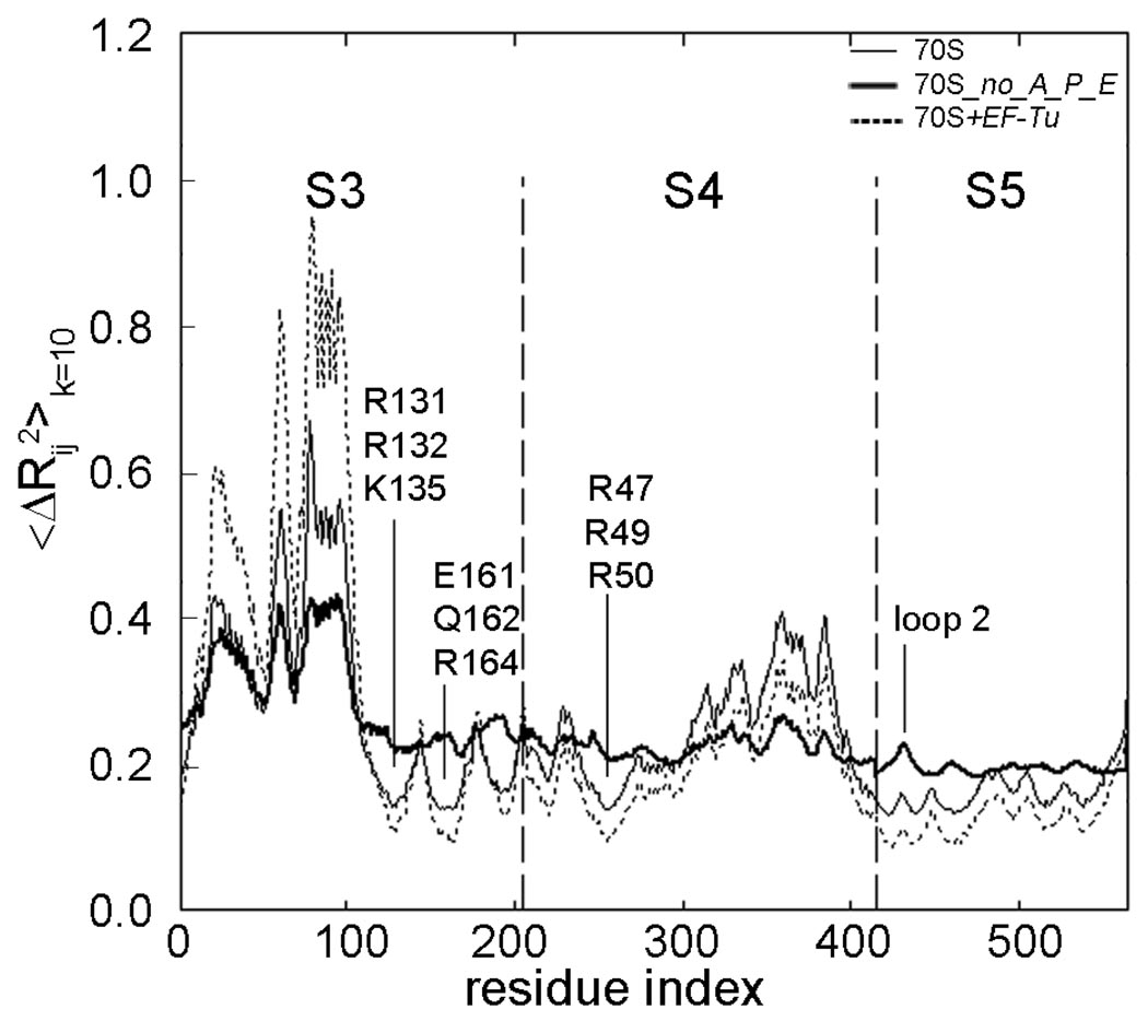
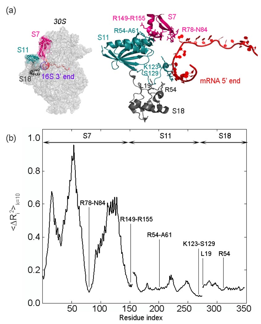
The protein-synthesizing ribosome undergoes large motions to effect the translocation of tRNAs and mRNA; here, the domain motions of this system are explored with a coarse-grained elastic network model using normal mode analysis. Crystal structures are used to construct various model systems of the 70S complex with/without tRNA, elongation factor Tu and the ribosomal proteins. Computed motions reveal the well-known ratchet-like rotational motion of the large subunits, as well as the head rotation of the small subunit and the high flexibility of the L1 and L7/L12 stalks, even in the absence of ribosomal proteins. This result indicates that these experimentally observed motions during translocation are inherently controlled by the ribosomal shape and only partially dependent upon GTP hydrolysis. Normal mode analysis further reveals the mobility of A- and P-tRNAs to increase in the absence of the E-tRNA. In addition, the dynamics of the E-tRNA is affected by the absence of the ribosomal protein L1. The mRNA in the entrance tunnel interacts directly with helicase proteins S3 and S4, which constrain the mRNA in a clamp-like fashion, as well as with protein S5, which likely orients the mRNA to ensure correct translation. The ribosomal proteins S7, S11 and S18 may also be involved in assuring translation fidelity by constraining the mRNA at the exit site of the channel. The mRNA also interacts with the 16S 3' end forming the Shine-Dalgarno complex at the initiation step; the 3' end may act as a 'hook' to reel in the mRNA to facilitate its exit.


Figures

Figure 1
Figure 1
Functionally important regions of the 70S ribosome complex (panel a). The small 30S subunit shown in transparent blue (panel a and b) contains the mRNA binding region, as well as its entrance and exit channels, together with the decoding center residues A1492 and A1493 residing on helix 44, at the interface of two subunits. The tRNA binding sites A-, P- and E- are indicated (panel a).The large 50S subunit in gray (panel a) has two prominent mobile stalks L1 and L7/L12, involved in controlling the tRNA exit and tRNA entrance, respectively. Extended ribosomal protein L9 of 50S is removed in some simulations to prevent its large dominant motions in normal mode analysis. The 30S subunit head has been reported to rotate during subunit association, tRNA selection and translocation, and this same type of rotation was also found in empty 70S E. coli ribosome crystal structure. The ribosomal proteins studied and the3’ end of 16S rRNA of the small subunit are shown in cartoon representation (panel b). All molecular graphics were prepared using PyMol.
Figure 2
Figure 2
Ribbon diagrams of some models for the 30S subunit, rRNA is represented in blue and ribosomal proteins in dark blue and for the 50S subunit, its rRNA is in grey and the ribosomal proteins in dark grey. (a) 70S and its complex with the A-, P- and E-tRNAs, together with the mRNA (b) the same 70S ribosome but also with EF-Tu and tRNA complex present in the hybrid A/T-site on 30S, and (c) rRNA of 70S and its complex with the A-, P-, E-tRNAs and mRNA, but without the proteins. PDB codes for the 30S and 50S subunits and EF-Tu are 1JGO, 1GIY and 1MJ1, respectively.
Figure 3
Figure 3
Displacement vectors between alternative conformations of different model systems for slow modes. One alternative displacement direction is shown with the parts of the structure in different colored blue/dark blue (30S rRNA/ proteins), gray/ dark gray (50S rRNA/ proteins) and magenta (EF-Tu) arrows. For clarity, the displacements of tRNAs are not shown.
Figure 4
Figure 4
Cumulative mean-square fluctuations over 10 slowest modes 〈ΔRi2k=10 for the (a) E-tRNA, (b) 23S rRNA region of L1 stalk in models 70S and 70S_no_L1, and (c) A- and P-tRNAs in models 70S and 70S_no_E. The motions are scaled between 0 and 1 for clarity.
Figure 5
Figure 5
Cumulative mean-square fluctuations over 10 slowest modes 〈ΔRi2k=10 for the regions (a) h44 and (b) H69 in models 70S, 70S+EF-Tu and 70S_no_A_P_E. The fluctuations are scaled between 0 and 1 for clarity.
Figure 6
Figure 6
(a) The mRNA (red) 3’ end surrounded by ribosomal proteins S3 (blue), S4 (orange) and S5 (green) and its 5’ end interacting with 16S 3’ end (purple). Residues reported to be important for the helicase activity and the translation fidelity of the ribosome,, are shown on the S3, S4 and S5. (b) Mean-square fluctuations between S3, S4, S5 proteins and the closest nucleotide A27 on mRNA cumulatively over the slowest 10 modes. Cumulative mean-square fluctuations summed over the slowest 10 modes 〈ΔRi2k=10 are shown for (c) mRNA and (d) the 3’ end of the 16S rRNA. It should be noted that all mean-square fluctuation values are relative and not scaled to match experimental B-factors. (e) Comparison of mRNA strands observed in various crystal structures; color codes for pdb files: 1JGO (blue), 2B64 (orange), 2J00 (green), 1YL4 (purple), 2HGR (black). When the different ribosome crystal structures (color coded as mentioned above) are compared, it is noteworthy that the side chains of the residues functioning in the helicase activity are very mobile.
Figure 7
Figure 7
Mean-square distance fluctuations 〈ΔRij2k=10 based on the cumulative sum of the slowest ten modes plotted for S3, S4, S5 between the closest mRNA nucleotide A27 for the model systems 70S, 70S_no_A_P_E and 70S+EF-Tu. The curves have been normalized and the motions are rescaled for purposes of comparison among the different parts of the structure.
Figure 8
Figure 8
(a) The 5’ end of the mRNA (red) is surrounded by ribosomal proteins S7 (magenta), S11 (turquoise) and S18 (gray) at the exit channel, shown in two views. The important residues reported in various studies, ,, are shown on the right in stick form. (b) Cumulative mean-square fluctuations summed over the slowest 10 modes 〈ΔRi2k=10 plotted for S7, S11 and S18, and rescaled between 0 and 1 . It is clear that the residues indicated in (a) and (b) surrounding the mRNA undergo extremely small fluctuations in ten slowest most important modes.

Similar articles

Cited by

References

    1. Agrawal RK, Penczek P, Grassucci RA, Li Y, Leith A, Nierhaus KH, Frank J. Direct visualization of A-, P-, and E-site transfer RNAs in the Escherichia coli ribosome. Science. 1996;271:1000–1002. - PubMed
    1. Yusupov MM, Yusupova GZ, Baucom A, Lieberman K, Earnest TN, Cate JH, Noller HF. Crystal structure of the ribosome at 5.5 A resolution. Science. 2001;292:883–896. - PubMed
    1. Nissen P, Hansen J, Ban N, Moore PB, Steitz TA. The structural basis of ribosome activity in peptide bond synthesis. Science. 2000;289:920–930. - PubMed
    1. Agrawal RK, Penczek P, Grassucci RA, Frank J. Visualization of elongation factor G on the Escherichia coli 70S ribosome: The mechanism of translocation. Proc Natl Acad Sci U.S.A. 1998;95:6134–6138. - PMC - PubMed
    1. Agrawal RK, Heagle AB, Penczek P, Grassucci RA, Frank J. EF-G-dependent GTP hydrolysis induces translocation accompanied by large conformational changes in the 70S ribosome. Nat. Struct. Biol. 1999;6:643–647. - PubMed

Publication types

LinkOut - more resources