Review
. 2009 Jan;6(1):16-26.
doi: 10.1038/ncpcardio1397. Epub 2008 Nov 25.

Hemodynamic shear stress and the endothelium in cardiovascular pathophysiology

Affiliations
Review

Hemodynamic shear stress and the endothelium in cardiovascular pathophysiology

Peter F Davies. Nat Clin Pract Cardiovasc Med. 2009 Jan.

Abstract

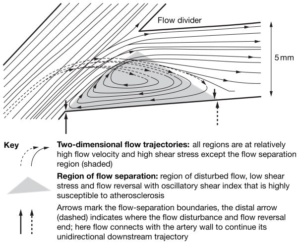
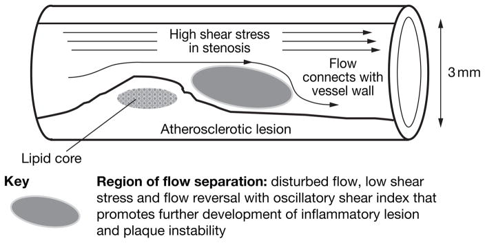
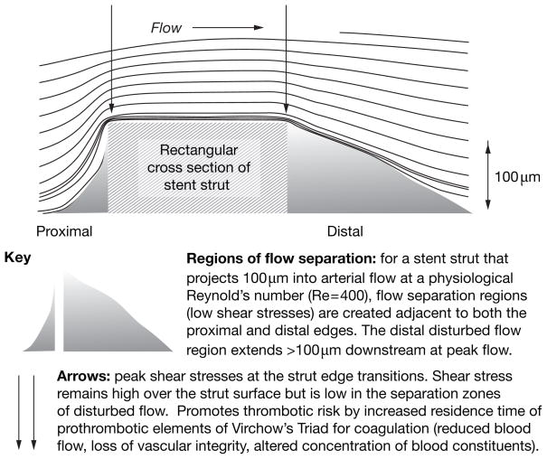
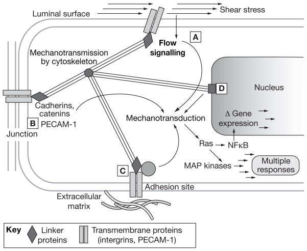
Endothelium lining the cardiovascular system is highly sensitive to hemodynamic shear stresses that act at the vessel luminal surface in the direction of blood flow. Physiological variations of shear stress regulate acute changes in vascular diameter and when sustained induce slow, adaptive, structural-wall remodeling. Both processes are endothelium-dependent and are systemically and regionally compromised by hyperlipidemia, hypertension, diabetes and inflammatory disorders. Shear stress spans a range of spatiotemporal scales and contributes to regional and focal heterogeneity of endothelial gene expression, which is important in vascular pathology. Regions of flow disturbances near arterial branches, bifurcations and curvatures result in complex spatiotemporal shear stresses and their characteristics can predict atherosclerosis susceptibility. Changes in local artery geometry during atherogenesis further modify shear stress characteristics at the endothelium. Intravascular devices can also influence flow-mediated endothelial responses. Endothelial flow-induced responses include a cell-signaling repertoire, collectively known as mechanotransduction, that ranges from instantaneous ion fluxes and biochemical pathways to gene and protein expression. A spatially decentralized mechanism of endothelial mechanotransduction is dominant, in which deformation at the cell surface induced by shear stress is transmitted as cytoskeletal tension changes to sites that are mechanically coupled to the cytoskeleton. A single shear stress mechanotransducer is unlikely to exist; rather, mechanotransduction occurs at multiple subcellular locations.


Conflict of interest statement

Competing interests

The author declared no competing interests.

Figures

Figure 1
Figure 1
Flow separations at an arterial branch can predispose or contribute to pathogenesis. Flow separation and the flow disturbance that occurs in the separated region are proatherogenic—complex, transient vortices form and dissipate (but not completely) throughout each heartbeat. The primary characteristics of disturbed flow are low average shear stress, constantly changing gradients of shear stress, oscillatory flow (and shear stress) because of flow reversal, and multifrequency, multidirectional, secondary flows. High shear stress protects against atherosclerosis as long as it remains below levels that detach the endothelium (estimated >40 N/m2 ; rare). In pulsatile flow within arteries, the separation region expands and contracts with the cardiac cycle. Excellent illustrations of computational imaging of human arterial hemodynamics can be viewed online.
Figure 2
Figure 2
Flow separations at a stenosis that predispose to or contribute to pathogenesis.
Figure 3
Figure 3
Flow separations around a stent strut that predispose to or contribute to pathogenesis.
Figure 4
Figure 4
The decentralized model of endothelial mechanotransduction by shear stress. The cytoskeleton has a central role in the transmission of tension changes throughout the cell. (A) Direct signaling can occur through deformation of the luminal surface, possibly via the glycocalyx. Examples include localized activation of potassium, sodium and calcium ion channels, phospholipase activity leading to calcium signaling, G-protein activation and caveolar signaling. (B) Mechanotransduction is also mediated via junctional signaling: that is, the transmission of forces to intercellular junction protein complexes via the cortical and/or filamentous cytoskeleton. VEGFR2 located at the luminal surface (see A) or near the junction (see B) can associate with VE-cadherin, β-catenin, and phosphatidylinositol 3 kinase to phosphorylate Akt and the primary transmembrane protein at this location, PECAM-1. (C) Cytoskeletal forces are also transmitted to adhesion sites. Transmembrane integrins bound to the extracellular matrix serve as a focus for deformation. This deformation results in autophosphorylation of FAK, which binds the SH2 domain of c-Src, a kinase family that phosphorylates paxillin and p130cas and leads to integrin-dependent activation of MAP kinases via Ras GTPase. A second parallel integrin-mediated pathway involves the activation of Shc, which binds Src family kinases through SH2 domains. Shc phosphorylation leads to Ras-MAP kinase activity. Ras releases the trans-acting NFκB from its cytosolic inhibitor, and thus enables its translocation to the nucleus where it binds to the promoters of multiple target genes. A third integrin-mediated pathway involves rhoA activation, which profoundly influences actin assembly and, therefore, transmission of mechanical stimuli. The multiple roles of small GTPases in mechanotransduction are reviewed in depth by Tzima. (D) Nuclear deformation is also likely to result in mechanically induced signaling, possibly via lamins in the nuclear membrane. Other possible direct effects include macromolecular conformation changes that are relevant to gene regulation. Of note, the locations are based on direct or indirect experimental evidence, are not mutually exclusive, and are probably interconnected. Permission obtained from Cambridge University Press © Davies PF and Helmke BP (2008) Endothelial Mechanotransduction. In Cellular Mechanotransduction: Diverse Perspectives from Molecules to Tissue (Eds Mofrad RK and Kamm RD). Abbreviations: Akt, protein kinase B; c-Src, tyrosine-protein kinase; FAK, focal adhesion kinase; MAP, mitogen-activated protein; NFκB, nuclear factor κB; p130Cas, Crk-associated substrate; PECAM-1, platelet endothelial cell adhesion molecule 1; Ras, small GTPase; Rho, small GTPase related to Ras; SH2, Src homology 2 domain; Shc, SH2-combining adaptor protein; VE-cadherin, vascular endothelial cadherin; VEGFR2, vascular endothelial growth factor receptor 2.

References

    1. Pohl U, et al. Crucial role of endothelium in the vasodilator response to increased flow in vivo. Hypertension. 1986;8:37–44. - PubMed
    1. Moncada S. Adventures in vascular biology: a tale of two mediators. Philos Trans R Soc Lond B Biol Sci. 2006;361:735–759. - PMC - PubMed
    1. Corson MA, et al. Phosphorylation of endothelial nitric oxide synthase in response to fluid shear stress. Circ Res. 1996;79:984–991. - PubMed
    1. Griffith TM. Endothelial control of vascular tone by nitric oxide and gap junctions: a haemodynamic perspective. Biorheology. 2002;39:307–318. - PubMed
    1. Davies PF. Flow-mediated endothelial mechanotransduction. Physiol Rev. 1995;75:519–560. - PMC - PubMed

Publication types

MeSH terms