

Interictal dysfunction of a brainstem descending modulatory center in migraine patients
- PMID: 19030105
- PMCID: PMC2582961
- DOI: 10.1371/journal.pone.0003799
Interictal dysfunction of a brainstem descending modulatory center in migraine patients
Abstract
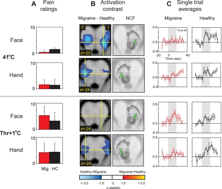
Background: The brainstem contains descending circuitry that can modulate nociceptive processing (neural signals associated with pain) in the dorsal horn of the spinal cord and the medullary dorsal horn. In migraineurs, abnormal brainstem function during attacks suggest that dysfunction of descending modulation may facilitate migraine attacks, either by reducing descending inhibition or increasing facilitation. To determine whether a brainstem dysfunction could play a role in facilitating migraine attacks, we measured brainstem function in migraineurs when they were not having an attack (i.e. the interictal phase).
Methods and findings: Using fMRI (functional magnetic resonance imaging), we mapped brainstem activity to heat stimuli in 12 episodic migraine patients during the interictal phase. Separate scans were collected to measure responses to 41 degrees C and noxious heat (pain threshold+1 degrees C). Stimuli were either applied to the forehead on the affected side (as reported during an attack) or the dorsum of the hand. This was repeated in 12 age-gender-matched control subjects, and the side tested corresponded to that in the matched migraine patients. Nucleus cuneiformis (NCF), a component of brainstem pain modulatory circuits, appears to be hypofunctional in migraineurs. 3 out of the 4 thermal stimulus conditions showed significantly greater NCF activation in control subjects than the migraine patients.
Conclusions: Altered descending modulation has been postulated to contribute to migraine, leading to loss of inhibition or enhanced facilitation resulting in hyperexcitability of trigeminovascular neurons. NCF function could potentially serve as a diagnostic measure in migraine patients, even when not experiencing an attack. This has important implications for the evaluation of therapies for migraine.
Conflict of interest statement
Figures
Comment in
-
Insights into the pathophysiology of headache provided by recent functional imaging studies.Headache. 2010 Oct;50(9):1528-30. doi: 10.1111/j.1526-4610.2010.01762.x. Headache. 2010. PMID: 20958299 No abstract available.
References
-
- Rocca MA, Ceccarelli A, Falini A, Colombo B, Tortorella P, et al. Brain gray matter changes in migraine patients with T2-visible lesions: a 3-T MRI study. Stroke. 2006;37:1765–1770. - PubMed
-
- Welch KM, Nagesh V, Aurora SK, Gelman N. Periaqueductal gray matter dysfunction in migraine: cause or the burden of illness? Headache. 2001;41:629–637. - PubMed
-
- Weiller C, May A, Limmroth V, Juptner M, Kaube H, et al. Brain stem activation in spontaneous human migraine attacks. Nat Med. 1995;1:658–660. - PubMed
-
- Afridi SK, Giffin NJ, Kaube H, Friston KJ, Ward NS, et al. A positron emission tomographic study in spontaneous migraine. Arch Neurol. 2005;62:1270–1275. - PubMed
-
- Bahra A, Matharu MS, Buchel C, Frackowiak RS, Goadsby PJ. Brainstem activation specific to migraine headache. Lancet. 2001;357:1016–1017. - PubMed
Publication types
MeSH terms
Grants and funding
LinkOut - more resources
Full Text Sources
Medical
