. 2009 Apr;131(4):471-81.
doi: 10.1007/s00418-008-0541-5. Epub 2008 Nov 22.

Characterization of the inflammatory and fibrotic response in a mouse model of cardiac pressure overload

Affiliations

Characterization of the inflammatory and fibrotic response in a mouse model of cardiac pressure overload

Ying Xia et al. Histochem Cell Biol. 2009 Apr.

Abstract

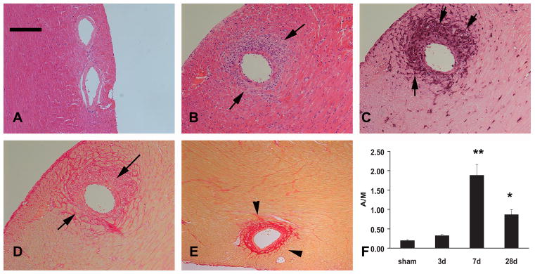
Myocardial fibrosis is an integral component of most cardiac pathologic conditions and contributes to the development of both systolic and diastolic dysfunction. Because of the availability of genetically manipulated animals, mouse models are essential for understanding the mechanisms involved in the pathogenesis of cardiac fibrosis. Accordingly, we characterized the inflammatory and fibrotic response in a mouse model of cardiac pressure overload due to transverse aortic constriction (TAC). Following TAC, mouse hearts exhibited induction of chemokines and proinflammatory cytokines, associated with macrophage, but not neutrophil, infiltration. Induction of inflammatory cytokines was followed by a late upregulation of transforming growth factor (TGF)-beta isoforms, activation of the Smad2/3 and Smad1/5 pathways, induction of matricellular proteins, and deposition of collagen. Inflammatory activity decreased after 28 days of TAC; at this timepoint established fibrosis was noted, accompanied by ventricular dilation and systolic dysfunction. Late induction of inhibitory mediators, such as TGF-beta, may play an essential role in the transition from inflammation to fibrosis by suppressing inflammatory gene synthesis while inducing matrix deposition. Our findings identify molecular mediators and pathways with a potential role in cardiac fibrosis laying the foundations for studies exploring the pathogenesis of fibrotic cardiac remodeling using genetically targeted mice.


Figures

Figure 1
Figure 1
Induction of proinflammatory cytokines in the pressure-overloaded myocardium. RPA analysis demonstrated upregulation of TNF-α (A), and IL-1β (B) mRNA after 3days of aortic banding (*p<0.05 vs. sham). TNF-α and IL-1β mRNA expression returned to sham levels after 7 days of TAC. IL-6 mRNA expression was not induced in the pressure-overloaded heart (C).
Figure 2
Figure 2
Chemokine upregulation in the pressure-overloaded mouse heart. RPA analysis demonstrated that mRNA expression of the CXC chemokines MIP-2 (A) and IP-10 (B) and the CC chemokine MCP-1 (C) was induced after 7 days of TAC (**p<0.01 vs. sham).
Figure 3
Figure 3
The time course of leukocyte infiltration in the pressure-overloaded mouse heart. Mac-2 immunohistochemistry identified macrophages in the heart after 3 days (A), 7 days (B) and 28 days (C) of TAC. Macrophage density peaked after 7 days of aortic banding (E, **p<0.01 vs. sham). Partial resolution of the macrophage infiltrate was noted after 28 days of aortic constriction (ˆp<0.05 vs. 7days). In contrast, only modest infiltration of the pressure overloaded myocardium with neutrophils (arrows) was noted after 3 days of TAC (D). The increase in neutrophil density did not reach statistical significance (E). Scale bar=100μm.
Figure 4
Figure 4
Induction of matricellular proteins and collagen deposition in the pressure-overloaded mouse heart. α-smooth muscle actin staining identified abundant spindle-shaped myofibroblasts in the pressure-overloaded myocardium after 7 days of TAC (A- arrows). Upregulation of the matricellular proteins osteopontin (OPN) and tenascin-C was noted in the fibrotic myocardium. OPN mRNA was induced after 3 days of aortic banding (B) (**p<0.01 vs. sham). Interstitial deposition of tenascin-C, a protein that is not expressed in the normal heart but is induced in remodeling tissues, peaked after 7 days (**p<0.01 vs. sham) (C, D - arrows) of aortic banding. Sirius red staining identifies the collagen network in the interstitial and perivascular space of the normal heart (arrowheads). After 7 days of aortic banding, expansion of the collagen-stained area was noted in both interstitial (arrows) and periarteriolar areas (arrowhead). Quantitative analysis demonstrated a significant increase of the area stained for collagen in all segments of the pressure-overloaded heart, peaking after 7 days of TAC (G, *p<0.05 vs. corresponding sham, **p<0.01 vs. corresponding sham). Symbols: LV, left ventricle; RV, Right ventricle. Scale bar=100 μm.
Figure 5
Figure 5
Cardiac pressure overload results in development of periarteriolar inflammation and fibrosis. A: H&E staining of a normal mouse heart demonstrates that intramyocardial arterioles have a thin adventitial layer. B-D: Serial section staining of the murine myocardium after 7 days of TAC for H&E (B), Mac-2 immunohistochemistry to identify macrophages (C) and sirius red to label the collagen network (D). Expansion of the arteriolar adventitia is noted (arrows), associated with macrophage infiltration (C – arrows) and deposition of collagen (D –arrows). After 28 days of aortic banding, the fibrotic arteriolar adventitia shows deposition of dense collagen fibers (E – arrowheads). F: The ratio of the adventitial to medial arteriolar area (A:M) was used to assess the extent of adventitial expansion. The A:M area significantly increases after 7-28 days of TAC (**p<0.01 vs. sham; *p<0.05 vs. sham). Scale bar=100μm
Figure 6
Figure 6
Induction of TGF-β isoforms and activation of TGF-β signaling pathways in the pressure overloaded mouse heart. RPA analysis demonstrated significant TGF-β1 (A), -β2 (B) and -β3 (C) mRNA upregulation after 7 days of aortic banding. Western blotting analysis showed a marked increase in the ratio of p-Smad2:Smad2 (D) and p-Smad1:Smad1 (E) after 7 days of TAC indicating activation of the Smad2/3 and Smad1/5 pathways.
Figure 7
Figure 7
Identification of microvascular endothelial cells in the pressure-overloaded myocardium using CD31 immunohistochemistry (A, sham; B, 3 days TAC; C, 28 days TAC). Quantitative analysis of microvascular density was performed in the subendocardial and subepicardial areas, where the vessels are seen in cross-section. Microvascular density significantly increased in all myocardial segments of the pressure-overloaded heart peaking after 3 days of TAC (D) (*p<0.05 vs. corresponding sham, **p<0.01 vs. corresponding sham, ˆp<0.05 vs corresponding LV epi). Symbols: LV epi, left ventricular subepicardial segment; LV endo, left ventricular subendocardial segment; RV, Right ventricle. Scale bar=60μm
Figure 8
Figure 8
Structural alterations in the pressure-overloaded mouse heart. A-C: Low magnification images of H&E-stained sections at the papillary muscle level (A, sham; B, 7 days TAC; C, 28days TAC) Scale bar=1mm. D-F M-mode echocardiographic images of the mouse heart (D, sham; E, 7 days TAC; F, 28 days TAC). Note that after 7 days of aortic banding the pressure-overloaded heart exhibits marked hypertrophy with normal chamber dimensions. LVEDD (white vertical line) and LVESD (white arrows) are comparable between sham mice (D) and animals undergoing 7 days of TAC (E). In contrast, left ventricular wall thickness (yellow arrows) is significantly increased after 7 days of banding. After 28 days of TAC significant ventricular dilation is noted accompanied by systolic dysfunction. Quantitative analysis of the echocardiographic findings is shown in Table 1.

References

    1. Beg AA. Endogenous ligands of Toll-like receptors: implications for regulating inflammatory and immune responses. Trends Immunol. 2002;23:509–512. - PubMed
    1. Berk BC, Fujiwara K, Lehoux S. ECM remodeling in hypertensive heart disease. J Clin Invest. 2007;117:568–575. - PMC - PubMed
    1. Brown RD, Ambler SK, Mitchell MD, Long CS. The cardiac fibroblast: therapeutic target in myocardial remodeling and failure. Annu Rev Pharmacol Toxicol. 2005;45:657–687. - PubMed
    1. Brown RD, Jones GM, Laird RE, Hudson P, Long CS. Cytokines regulate matrix metalloproteinases and migration in cardiac fibroblasts. Biochem Biophys Res Commun. 2007;362:200–205. - PMC - PubMed
    1. Bryant D, Becker L, Richardson J, Shelton J, Franco F, Peshock R, Thompson M, Giroir B. Cardiac failure in transgenic mice with myocardial expression of tumor necrosis factor-alpha. Circulation. 1998;97:1375–1381. - PubMed

Publication types

MeSH terms

LinkOut - more resources