. 2008 Nov 21;3(11):703-10.
doi: 10.1021/cb800185h.

Probing the function of heme distortion in the H-NOX family

Affiliations

Probing the function of heme distortion in the H-NOX family

Charles Olea et al. ACS Chem Biol. .

Abstract

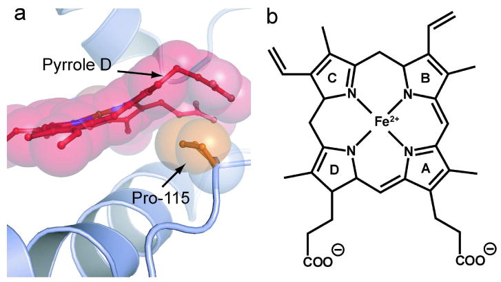
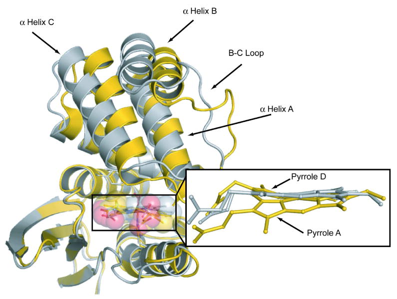
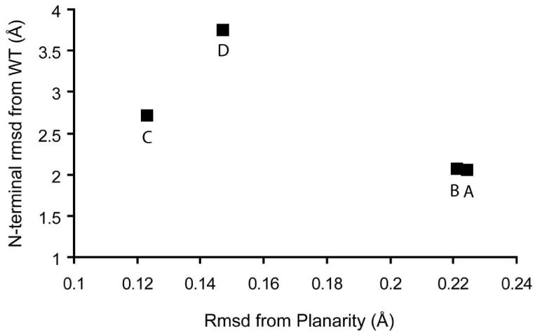
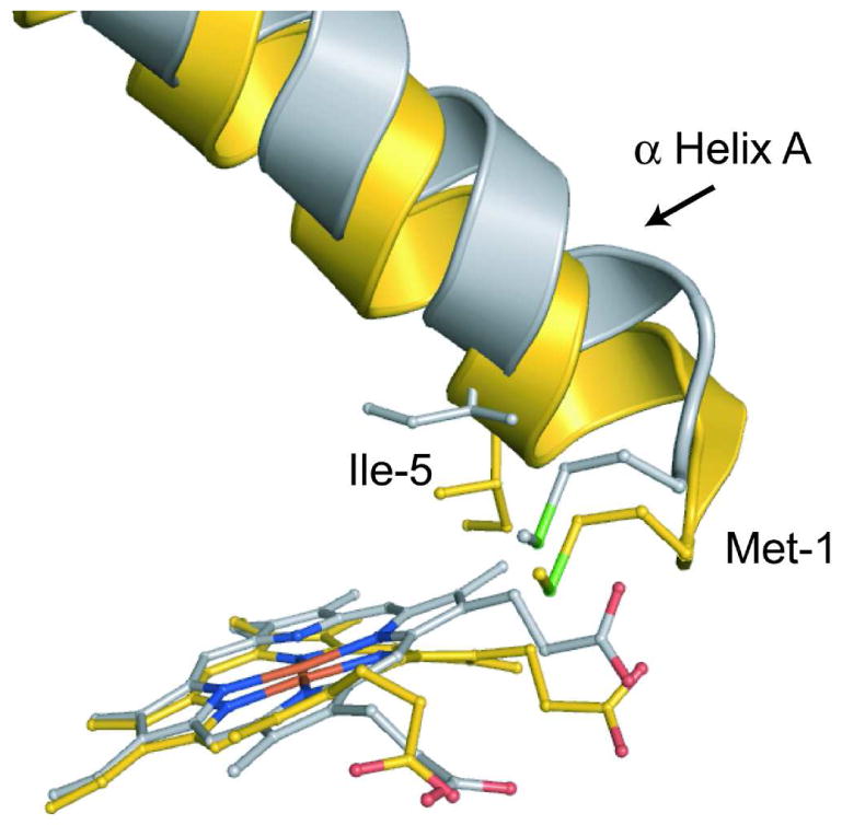
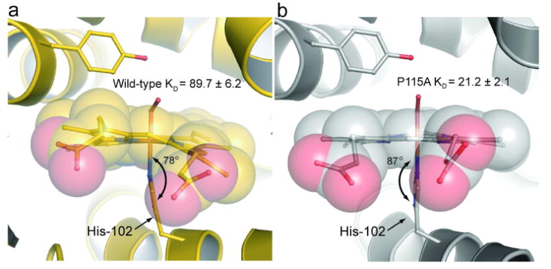
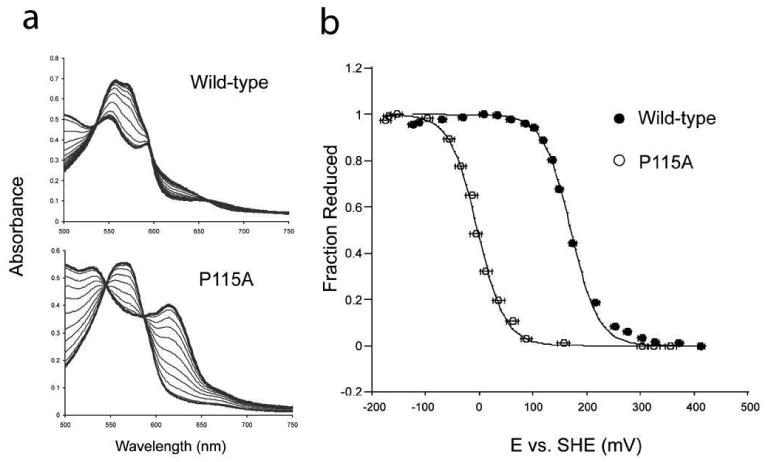
Hemoproteins carry out diverse functions utilizing a wide range of chemical reactivity while employing the same heme prosthetic group. It is clear from high-resolution crystal structures and biochemical studies that protein-bound hemes are not planar and adopt diverse conformations. The crystal structure of an H-NOX domain from Thermoanaerobacter tengcongensis (Tt H-NOX) contains the most distorted heme reported to date. In this study, Tt H-NOX was engineered to adopt a flatter heme by mutating proline 115, a conserved residue in the H-NOX family, to alanine. Decreasing heme distortion in Tt H-NOX increases affinity for oxygen and decreases the reduction potential of the heme iron. Additionally, flattening the heme is associated with significant shifts in the N-terminus of the protein. These results show a clear link between the heme conformation and Tt H-NOX structure and demonstrate that heme distortion is an important determinant for maintaining biochemical properties in H-NOX proteins.


Figures

Figure 1
Figure 1. Heme Distortion in wild-type Tt H-NOX
a) Shown is a ball-and-stick and space filling model of P115 and the surrounding heme environment of wild-type Tt H-NOX (15). The invariant P115 (orange) makes the largest contribution to heme (red) distortion in Tt H-NOX. P115 pushes up against pyrrole D, which causes a pronounced kink in the connected propionate group. Out-of-plane distortions of up to 2 Å are observed in Tt H-NOX. b) Heme prosthetic group with pyrrole groups A-D labeled.
Figure 2
Figure 2. Structural comparison of P115A with wild-type Tt H-NOX
The heme in P115A (silver) is flatter than wild-type (gold). Significant translations are observed in the N-terminal region of the heme-flattened P115A crystal structure. Shown are molecule A from the monoclinic crystal structure of wild-type H-NOX and molecule D from the P115A crystal structure.
Figure 3
Figure 3. N-terminal movement from wild-type Tt H-NOX (WT) vs. heme distortion in P115A
The N-terminal (residues 1–83) rms deviation (Å) was calculated and plotted vs. rms deviation (Å) from planarity for each of the 4 molecules in the asymmetric unit cell (A-D). Wild-type molecule A in the monoclinic space group was used for analysis.
Figure 4
Figure 4. The effect of heme flattening on the N-terminal domain
Shown is a comparison of P115A molecule D (silver) and wild-type monoclinic Tt H-NOX molecule A (gold) heme/N-terminal interface. The planar heme makes new contacts with Met1 and Ile5 of α helix A, which shifts the helix away from the C-terminal domain. Shifting of α helix A, along with the rest of the N-terminal region causes shifts over 4.9 Å.
Figure 5
Figure 5. Comparison of the iron-histidine bond geometry
Shown are the iron-histidine tilts of a) wild-type Tt H-NOX (gold) and b) P115A (silver). The least squares plane of the heme was calculated using the 4 pyrrole nitrogens of the heme (other atoms were excluded due to the high degree of distortion) and the five imidazole ring atoms of H102 using MOLEMAN2 (22). Wild-type has a tilt of 78° whereas P115A has a tilt of 87°.
Figure 6
Figure 6. Reduction potential of wild-type Tt H-NOX and P115A
a) Shown are the titration spectra for wild-type Tt H-NOX and P115A. b) Titration curves for wild-type (•) and P115A (o). The reduction potentials of P115A and wild-type Tt H-NOX were determined against the standard hydrogen electrode (SHE). The ratio of reduced Fe2+ to oxidized Fe3+ heme was measured based on their α/β maximum at approximately 557 (reduced) nm for wild-type. The difference absorbance of the α/β maximum for reduced and the α/β minimum for oxidized was used to calculate the fraction reduced for P115A. The voltage against the SHE was measured for both oxidative and reductive titrations of wild-type and P115A. Error bars represent the standard error.

Comment in

  • A twist on heme signaling.
    Spiro T. Spiro T. ACS Chem Biol. 2008 Nov 21;3(11):673-5. doi: 10.1021/cb800269h. ACS Chem Biol. 2008. PMID: 19032089 Free PMC article.

References

    1. Shelnutt JA, Song XZ, Ma JG, Jia SL, Jentzen W, Medforth CJ. Nonplanar porphyrins and their significance in proteins. Chem Soc Rev. 1998;27:31–41.
    1. Zbylut SD, Kincaid JR. Resonance Raman evidence for protein-induced out-of-plane distortion of the heme prosthetic group of mammalian lactoperoxidase. J Amer Chem Soc. 2002;124:6751–6758. - PubMed
    1. Li D, Stuehr DJ, Yeh SR, Rousseau DL. Heme distortion modulated by ligand-protein interactions in inducible nitric oxide synthase. J Biol Chem. 2004;279:26489–26499. - PubMed
    1. Ravikanth M, Chandrashekar TK. Nonplanar porphyrins and their biological relevance - ground and excited-state dynamics. Coordination Chemistry. 1995;82:105–188.
    1. Weichsel A, Andersen JF, Roberts SA, Montfort WR. Nitric oxide binding to nitrophorin 4 induces complete distal pocket burial. Nature Structural Biology. 2000;7:551–554. - PubMed

Publication types