Comparative Study
. 2008;10(6):R137.
doi: 10.1186/ar2560. Epub 2008 Nov 25.

Identification of possible candidate genes regulating Sjögren's syndrome-associated autoimmunity: a potential role for TNFSF4 in autoimmune exocrinopathy

Affiliations
Comparative Study

Identification of possible candidate genes regulating Sjögren's syndrome-associated autoimmunity: a potential role for TNFSF4 in autoimmune exocrinopathy

Cuong Q Nguyen et al. Arthritis Res Ther. 2008.

Abstract

Introduction: Sjögren syndrome (SjS) is a systemic autoimmune disease in which an immunological attack primarily against the salivary and lacrimal glands results in the loss of acinar cell tissue and function, leading to stomatitis sicca and keratoconjunctivitis sicca. In recent years, two genetic regions, one on chromosome 1 (designated autoimmune exocrinopathy 2 or Aec2) and the second on chromosome 3 (designated autoimmune exocrinopathy 1 or Aec1) derived from nonobese diabetic (NOD) mice, have been shown to be necessary and sufficient to replicate SjS-like disease in nonsusceptible C57BL/6 mice.

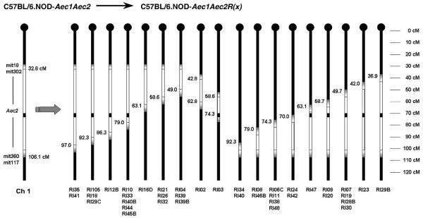
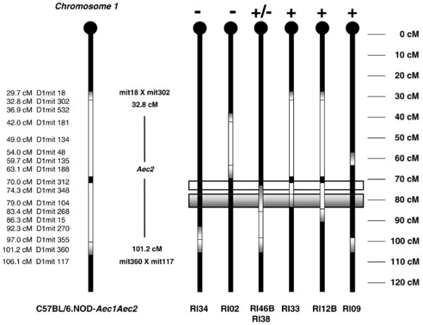
Methods: Starting with the SjS-susceptible C57BL/6-derived mouse, referred to as C57BL/6.NOD-Aec1Aec2, we generated a large set of recombinant inbred (RI) lines containing portions of Aec2 as a means of identifying more precisely the genetic elements of chromosome 1 responsible for disease development.

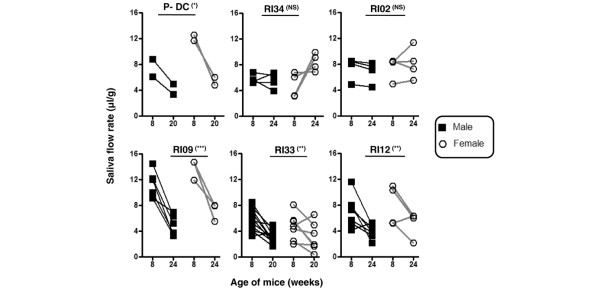
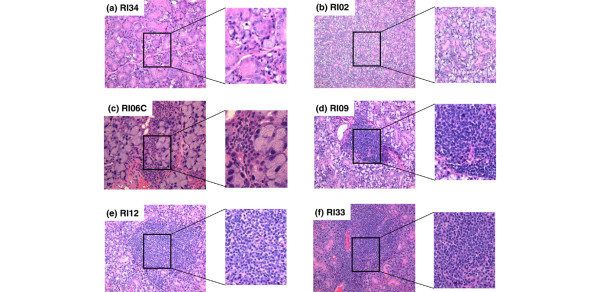
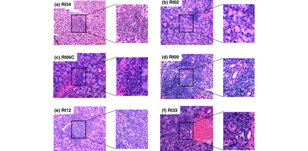
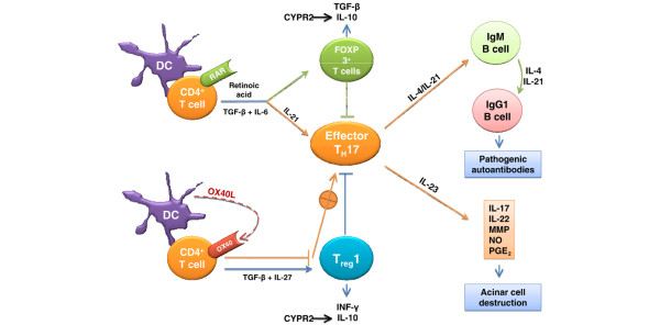
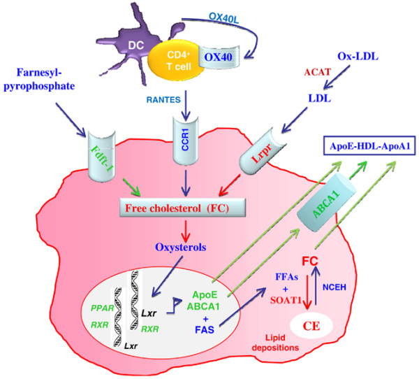
Results: Disease profiling of these RI lines has revealed that the SjS susceptibility genes of Aec2 lie within a region located at approximately 79 +/- 5 cM distal to the centromere, as defined by microsatellite markers. This chromosomal region contains several sets of genes known to correlate with various immunopathological features of SjS as well as disease susceptibility genes for both type 1 diabetes and systemic lupus erythematosus in mice. One gene in particular, tumor necrosis factor (ligand) superfamily member 4 (or Ox40 ligand), encoding a product whose biological functions correlate with both physiological homeostasis and immune regulations, could be a potential candidate SjS susceptibility gene.

Conclusions: These new RI lines represent the first step not only in fine mapping SjS susceptibility loci but also in identifying potential candidate SjS susceptibility genes. Identification of possible candidate genes permits construction of models describing underlying molecular pathogenic mechanisms in this model of SjS and establishes a basis for construction of specific gene knockout mice.


Figures

Figure 1
Figure 1
Map of chromosome 1 crossover points in C57BL/6.NOD-Aec1Aec2R(n) recombinant inbred (RI) mice. Thirty-nine RI lines are aligned to show the points of their individual crossovers in the Aec2 region of chromosome 1, as determined by D1mit microsatellite markers. Crossover frequencies are higher at approximately 49.7, 74.3, and 79.0 cM but are not considered hotspots for chromosomal 1 crossovers (NS: Not significant, * = p < 0.05, ** = p < 0.01, and *** = p < 0.001).
Figure 2
Figure 2
Differences in temporal loss of secretory function in various C57BL/6.NOD-Aec1Aec2R(n) mice. Male and female sibling mice of parental C57BL/6.NOD-Aec1Aec2 (P-DC) and C57BL/6.NOD-Aec1Aec2R(n) mice were injected with isoproterenol/pilocarpine, first at 8 weeks of age and then at 20 or 24 weeks of age, to stimulate saliva secretion. Saliva was collected from each mouse for 10 minutes starting 1 minute after injection of the secretagogue. The volume of each sample was measured and standardized relative to the weight of the mouse. Temporal reductions in saliva secretions, a marker for onset of clinical disease, were used to identify genetic regions containing genes necessary for development of salivary gland dysfunction and Sjögren syndrome. NS, not significant; RI, recombinant inbred.
Figure 3
Figure 3
Histological characterization of sialadenitis of male and female C57BL/6.NOD-Aec1Aec2R(n) mice. Submandibular glands were freshly explanted from male and female C57BL/6.NOD-Aec1Aec2R(n) mice euthanized at 20 or 24 weeks of age. The glands were fixed in 10% formalin, embedded in paraffin, and sectioned and stained with hematoxylin and eosin (H&E) dye. Representative H&E-stained histological sections of submandibular glands of selected recombinant inbred (RI) lines are presented: (a) RI34, (b) RI02, (c) RI06C, (d) RI09, (e) RI12, and (f) RI33. Original images were taken at × 100 magnification, with inserts expanded to show structural detail.
Figure 4
Figure 4
Histological characterization of dacryoadenitis of male and female C57BL/6.NOD-Aec1Aec2R(n) mice. Submandibular and lacrimal glands were freshly explanted from male and female C57BL/6.NOD-Aec1Aec2R(n) mice euthanized at 20 or 24 weeks of age. The glands were fixed in 10% formalin, embedded in paraffin, and sectioned and stained with hematoxylin and eosin (H&E) dye. Representative H&E-stained histological sections of lacrimal glands of selected recombinant inbred (RI) lines are presented: (a) RI34, (b) RI02, (c) RI06C, (d) RI09, (e) RI12, and (f) RI33. Original images were taken at × 100 magnification, with inserts expanded to show structural detail.
Figure 5
Figure 5
Detection of anti-nuclear autoantibodies in sera of C57BL/6.NOD-Aec1RAec2R(n) mice. Serum samples obtained from C57BL/6.NOD-Aec1Aec2R(n) mice were diluted 1:40 and incubated with HEp-2 fixed substrate slides for 30 minutes at 25°C in a humidified chamber. The slides were then developed with Alexa 594-conjugated goat anti-mouse IgG and viewed by fluorescence microscopy at × 100 magnification. Examples of speckled/homogenous staining of the nucleus (left panel), cytoplasmic/nuclear membrane staining (left center panel), speckled/cytoplasmic staining (right center panel), and cytoplasmic staining (right panel) were observed. The numbers of individual sera tested and the percentages of positive sera from a sampling of recombinant inbred (RI) lines exhibiting each of the patterns are listed. §Number of mice showing positive staining pattern over total. *Percentage of mice showing positive staining pattern. Aec, autoimmune exocrinopathy.
Figure 6
Figure 6
Redefining the boundaries for the Aec2 Sjögren Syndrome (SjS) susceptibility genetic locus. Based on the disease profiling of the C57BL/6.NOD-Aec1Aec2R(n) recombinant inbred (RI) lines, the boundaries of the Aec2 genetic region containing SjS susceptibility genes have been temporarily reset to position 79 ± 5 cM of chromosome 1 (shaded gray gradient rectangular box). Possible quantitative trait loci genes may reside a few centimorgans centromeric to this region (unshaded rectangular box).
Figure 7
Figure 7
Proposed model for how OX40L:OX40 promotes autoimmunity in Sjögren syndrome-like disease of C57BL/6.NOD-Aec1Aec2 mice. Cellular interactions involved in the development of an autoimmune response against the salivary and lacrimal glands leading to loss of acinar tissue are presented. DC, dendritic cell; IL, interleukin; INF-γ, interferon-gamma; MMP, matrix metalloproteinase; NO, nitric oxide, PGE2, prostaglandin E2; TGF-β, transforming growth factor-beta; Treg, regulatory T (cell).
Figure 8
Figure 8
Proposed genetic predisposition for dysregulated homeostasis/transport of lipid, lipoprotein, cholesterol, and fatty acid metabolism leading to lipid depositions in the salivary and lacrimal glands of C57BL/6.NOD-Aec1Aec2 mice and Sjögren syndrome patients. Accumulation of free cholesterols (FCs) inside the cells resulted from increased uptake of low- and high-density lipid receptors. In addition, impairment of ABCA1 membrane transporter leads to the accumulation of cholesteryl esters (CEs) metabolized by sterol O-acyltransferase-1 (SOAT-1) using FCs and free fatty acids (FFAs). ABCA1, ATP-binding cassette, subfamily A [ABC1] member 1; ACAT, acyl-coenzyme A: cholesterol acyltransferase; ApoE, apolipoprotein E; DC, dendritic cell; Fdft-1, farnesyl diphosphate farnesyl transferase-1; HDL, high-density lipid; LDL, low-density lipid; Lrpr, low-density lipid-related protein receptor; NCEH, neutral cholesterol esters hydrolase; Ox-LDL, oxidized low-density lipid; PPAR, peroxisome proliferator activated receptor; RANTES, regulated on activation normal T cell expressed and secreted; RXR, retinoid × receptor. Adapted from [48].

Comment in

References

    1. Fox RI, Kang HI. Pathogenesis of Sjögren's syndrome. Rheum Dis Clin North Am. 1992;18:517–538. - PubMed
    1. Hansen A, Lipsky PE, Dorner T. New concepts in the pathogenesis of Sjogren syndrome: many questions, fewer answers. Curr Opin Rheumatol. 2003;15:563–570. doi: 10.1097/00002281-200309000-00007. - DOI - PubMed
    1. Jonsson R, Haga HJ, Gordon TP. Current concepts on diagnosis, autoantibodies and therapy in Sjögren's syndrome. Scand J Rheumatol. 2000;29:341–348. doi: 10.1080/030097400447525. - DOI - PubMed
    1. Nguyen CQ, Cha SR, Peck AB. Sjögren's syndrome (SjS)-like disease of mice: the importance of B lymphocytes and autoantibodies. Front Biosci. 2007;12:1767–1789. doi: 10.2741/2187. - DOI - PubMed
    1. Makino S, Kunimoto K, Muraoka Y, Mizushima Y, Katagiri K, Tochino Y. Breeding of a non-obese, diabetic strain of mice. Jikken Dobutsu. 1980;29:1–13. - PubMed

Publication types

MeSH terms