. 2008 Nov 25:3:48.
doi: 10.1186/1745-6150-3-48.

Functional insight into Maelstrom in the germline piRNA pathway: a unique domain homologous to the DnaQ-H 3'-5' exonuclease, its lineage-specific expansion/loss and evolutionarily active site switch

Affiliations

Functional insight into Maelstrom in the germline piRNA pathway: a unique domain homologous to the DnaQ-H 3'-5' exonuclease, its lineage-specific expansion/loss and evolutionarily active site switch

Dapeng Zhang et al. Biol Direct. .

Abstract

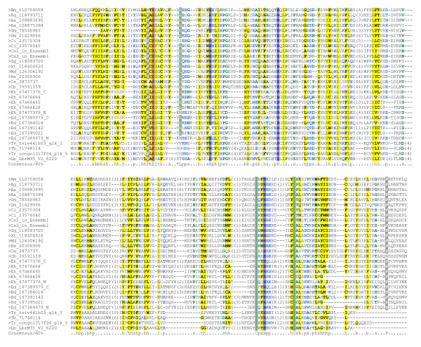
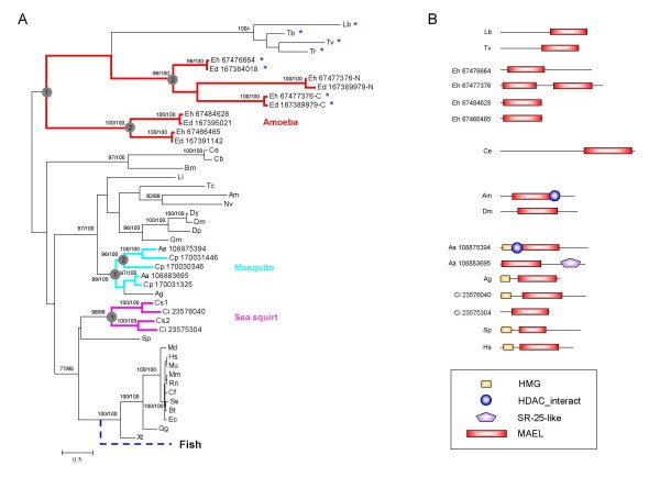
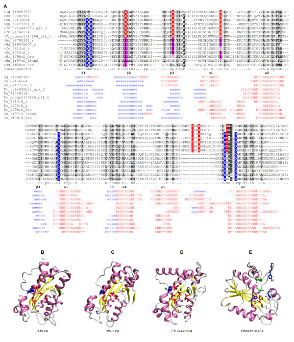
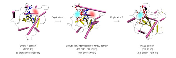
Maelstrom (MAEL) plays a crucial role in a recently-discovered piRNA pathway; however its specific function remains unknown. Here a novel MAEL-specific domain characterized by a set of conserved residues (Glu-His-His-Cys-His-Cys, EHHCHC) was identified in a broad range of species including vertebrates, sea squirts, insects, nematodes, and protists. It exhibits ancient lineage-specific expansions in several species, however, appears to be lost in all examined teleost fish species. Functional involvement of MAEL domains in DNA- and RNA-related processes was further revealed by its association with HMG, SR-25-like and HDAC_interact domains. A distant similarity to the DnaQ-H 3'-5' exonuclease family with the RNase H fold was discovered based on the evidence that all MAEL domains adopt the canonical RNase H fold; and several protist MAEL domains contain the conserved 3'-5' exonuclease active site residues (Asp-Glu-Asp-His-Asp, DEDHD). This evolutionary link together with structural examinations leads to a hypothesis that MAEL domains may have a potential nuclease activity or RNA-binding ability that may be implicated in piRNA biogenesis. The observed transition of two sets of characteristic residues between the ancestral DnaQ-H and the descendent MAEL domains may suggest a new mode for protein function evolution called "active site switch", in which the protist MAEL homologues are the likely evolutionary intermediates due to harboring the specific characteristics of both 3'-5' exonuclease and MAEL domains.


Figures

Figure 1
Figure 1
Multiple sequence alignment of representatives of MAEL domain. The sequences are represented by an abbreviation of species name followed by database entry ID. The homologues of C. savignyi identified in Ensembl database are indicated by Cs1 and Cs2. The consensus in 80% of the sequences is shown below the alignment based on default amino acid classes in Chroma. The numbers in bracket are indicative of the excluded residues from sequences. For a complete multiple sequence alignment refer to additional file 1. Species name abbreviations: Aa, Aedes aegypti; Ag, Anopheles gambiae; Am, Apis mellifera; Cb, Caenorhabditis briggsae; Ce, Caenorhabditis elegans; Ci, Ciona intestinalis; Cs,Ciona savignyi; Dm, Drosophila melanogaster; Ed, Entamoeba dispar SAW760; Eh, Entamoeba histolytica; Gg, Gallus gallus; Gm, Glossina morsitans; Hs, Homo sapiens; Md, Monodelphis domestica; Lb, Leishmania braziliensis; Tb, Trypanosoma brucei TREU927; Tr, Trypanosoma congolense; Tv, Trypanosoma vivax; Xt, Xenopus tropicalis.
Figure 2
Figure 2
Phylogenetic relationship and domain architectures of MAEL proteins. (A) An unrooted phylogenetic tree was reconstructed using maximum likelihood (ML) analysis and Bayesian analysis. Single MAEL domains are represented by species names. The duplicated ones in the species of Aa, Ci, Cp, Ed, Eh are represented by species names following Genbank ID, whereas Cs domains are represented by Cs1 and Cs2. Branch length is proportional to estimated evolutionary change by PhyML program; the scale bar represents 0.5 substitution per site. Node supporting values greater than 75% from ML bootstrap analyses and Bayesian MCMCMC sampling are shown on the left and on the right of the slash, respectively. Lineage-specific expansions of MAEL domains in amoeba, mosquito and sea squirt are highlighted with different colors (red, turquoise blue and pink) and ancient duplication events were indicated by circled numbers. The loss of MAEL in teleost fish is indicated in blue dashed. Asterisk labeled MAEL domains are the ones containing both conserved EHHCHC and EDDHD residues (see following main text). (B) Domain architectures of representatives of the MAEL proteins were deduced through searching against Pfam and SMART domain databases and drawn approximately to scale. The domains shown are: HDAC_interact, named after Histone deacetylase (HDAC) interacting (SMART: SM00761); HMG, named after High Mobility Group (SMART: SM00398); SR-25-like (DUF1777, Pfam: PF08648). New species name abbreviations: Bm, Brugia malayi; Bt, Bos taurus; Cf, Canis familiaris; Cp, Culex pipiens quinquefasciatus; Dp, Drosophila pseudoobscura; Dy, Drosophila yakuba; Ec, Equus caballus; Lb, Leishmania braziliensis; Ll, Lutzomyia longipalpis; Mm, Mus musculus; Mu, Macaca mulatta; Nv, Nasonia vitripennis; Rn, Rattus norvegicus; Sp, Strongylocentrotus purpuratus; Ss, Sus scrofa; Tc, Tribolium castaneum. Other species name abbreviations refer to Figure 1 legend.
Figure 3
Figure 3
Sequence and structure similarity between MAEL and DnaQ-H domains. (A) Sequence and the secondary structure alignment of MAEL and DnaQ-H domains. Seven DnaQ-H domains are included and five domains have 3-D structures: Thermotaga maritime ε exonuclease (Tm ε, 2P1J:A), Escherichia coli ε exonuclease (Ec_ε, 1J53:A), Haemophilus influenzae oligoribonuclease (Hi_Orn, 1J9A:A), human Trex2 exonuclease (Hs_Trex2, 1Y97:A) and human 3’-5’ exoribonuclease (Hs_Exo, 1W0H:A). The conserved DnaQ-H specific residues (DEDHD) are highlighted with the blue background; whereas the conserved MAEL-specific residues (EHHCHC) are highlighted with the red background. The MAEL-specific residues (EHH) also exist at counterpart positions in some DnaQ-H domains, and were highlighted with pink background. Detailed secondary structures for MAEL domains and DnaQ-H domains are obtained from secondary structure predictions and the 3-D structures, respectively; they are shown below the alignment (h in red, α helix; e in blue, β sheet). The structural sequence alignment was established carefully by hand on the basis of CE-MC results, alignment in fold recognition, literature information, and predicted secondary structures. The numbers in bracket are indicative of the excluded residues from sequences. New species name abbreviations: Tm, Thermotaga maritime; Ac,Alteromonas macleodii; Ma,marine gamma proteobacterium. (B-E) NewCartoon diagrams for DnaQ-H domains (1J53:A and 1W0H:A) and the homology model of two MAEL domains (Eh 67476664 and chicken MAEL). The α helices are shown in pink, β sheets in yellow, and loops in white; Their spatial locations are labeled in 1J53:A. Strictly conserved DnaQ-H active site residues DEDHD of protist MAEL domain (Eh 67476664) are highlighted in licorice drawing with acidic residues (D and E) in red and basic His in blue. The MAEL-specific residues EHHCHC of chicken MAEL domain are highlighted with Glu in red, His in blue and Cys in orange.
Figure 4
Figure 4
A proposed "active site switch" mode for MAEL domain function evolution. The cartoon drawing of protein structure is shown. The α helices are shown in pink, β sheets in yellow and loops in white. The DnaQ-H specific residues (DEDHD) are highlighted in blue, whereas MAEL specific residues (EHHCHC) are highlighted in red. Red cloud and green cloud indicate DnaQ-H active site and MAEL-specific active site, respectively.

References

    1. Kotaja N, Sassone-Corsi P. The chromatoid body: a germ-cell-specific RNA-processing centre. Nat Rev Mol Cell Biol. 2007;8:85–90. doi: 10.1038/nrm2081. - DOI - PubMed
    1. Ikenishi K. Germ plasm in Caenorhabditis elegans, Drosophila and Xenopus. Dev Growth Differ. 1998;40:1–10. doi: 10.1046/j.1440-169X.1998.t01-4-00001.x. - DOI - PubMed
    1. Aravin AA, Lagos-Quintana M, Yalcin A, Zavolan M, Marks D, Snyder B, Gaasterland T, Meyer J, Tuschl T. The small RNA profile during Drosophila melanogaster development. Dev Cell. 2003;5:337–350. doi: 10.1016/S1534-5807(03)00228-4. - DOI - PubMed
    1. Vagin VV, Sigova A, Li C, Seitz H, Gvozdev V, Zamore PD. A distinct small RNA pathway silences selfish genetic elements in the germline. Science. 2006;313:320–324. doi: 10.1126/science.1129333. - DOI - PubMed
    1. Hartig JV, Tomari Y, Forstemann K. piRNAs–the ancient hunters of genome invaders. Genes Dev. 2007;21:1707–1713. doi: 10.1101/gad.1567007. - DOI - PubMed

Publication types