. 2009 Feb;482(1-2):83-95.
doi: 10.1016/j.abb.2008.11.009. Epub 2008 Nov 14.

Inhibition of early endosome fusion by Rab5-binding defective Ras interference 1 mutants

Affiliations

Inhibition of early endosome fusion by Rab5-binding defective Ras interference 1 mutants

Adriana Galvis et al. Arch Biochem Biophys. 2009 Feb.

Abstract

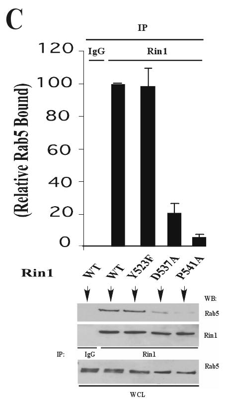
Rin1 has been shown to play an important role in endocytosis. In this study we demonstrated that depletion of Rin1 from the cytosol blocked the fusion reaction. More importantly, endosome fusion was rescued by the addition of Rin1 proteins depending on the presence of Rab5, and its effector EEA1. Furthermore, we found that Syntaxin 13, but not Syntaxin 7, was required by Rin1 to support endosome fusion. We also identified six mutations on the Vps9 domain of Rin1 that failed to rescue the fusion reaction. Two of them, Rin1: D537A and Rin1: Y561F mutants showed dramatic inhibitory effect on the fusion reaction, which correlate with their inability to properly activate Rab5 or to bind endosomal membranes. Taken together, our results suggest that specific residues on the Vsp9 domain of Rin1 are required for its interaction with Rab5, binding to the endosomal membranes and subsequent regulation of the fusion reaction.


Figures

Figure 1
Figure 1. Uptake and subcellular compartmentalization of biotin-EGF and avidin-β-galactosidase in Hela cells
Hela cells (6 × 10⁷) were incubated for 5 min at 37°C with 125I-biotin EGF (B-EGF, ■) or 125I-avidin-β-galactosidase (Av-Gal, ●) at 0.1 μg/ml and 2 mg/ml final concentration, respectively. The cells were then washed and fractionated. A membrane faction was mixed with 25 ml of Percoll (density 1.05 g/l) and centrifuged at 39,000 × g for 60 min. The gradients were collected from the bottom and analyzed for radioactivity and acid phosphatase activity (▲). Inset: Hela cells (1.5 × 10⁶) were incubated for 15 min at 37°C with different concentrations of 125I-Av-Gal (◆) or 125I-B-EGF (◇) as indicated in Material and Methods. The specific uptake is shown for each ligand concentration. Values are means ± SD, n=4.
Figure 2
Figure 2. Fusion between endosomes in intact cells
(A) Cells were incubated with Av-Gal for 1.5 min uptake, washed and chased for 1.5 min at 37°C. Cells were then washed, and then incubated at 4°C for 2 h with B-EGF. Endocytosis of EGF was started by incubation at 37°C and the amount of B-EGF associated with Av-Gal was quantitated at each time by solubilizing the cells in the presence of Biotin-BSA. Results are expressed as fluorescence units, which indicates the percentage of the immunocomplex formed after solubilization of vesicles in the absence of Biotin-BSA. Total was 68 ±1.2 fluorescence units and did not change significantly with the time of internalization. Values are means ± SD, n=4. (B) Outline of the method to determine an endosome-endosome fusion event in a cell-free system. Endosome fractions containing appropriate probes are co-incubated under fusogenic conditions (i.e., support vesicles fusion). These events (i.e., vesicle fusion) are evaluated by the Av-Gal∷B-EGF complex formation, which may be assayed in wells coated with anti-EGF antibodies and processed as described in Material and Methods.
Figure 3
Figure 3. Effect of temperature, time, cytosol, and energy on the fusion reaction
(A) Five-minute endosomes isolated from Hela cells containing B-EGF and Av-Gal were mixed in fusion buffer containing 2 mg/ml cytosol, incubated for 30 min at different temperatures in the presence of an ATP-regenerating (●, +ATP) or ATP-depleting (■, -ATP) systems and processed as described under Materials and Methods. Values are means ± SD, n=3. (B) Five-minute endosomes generated as described above were mixed in fusion buffer in the presence of 2 mg/ml cytosol containing an ATP-regenerating (●, +ATP) or ATP-depleting (■, -ATP) systems for the indicated time at 37°C, and processed as described above. Values are means ± SD, n=3. (C) Five-minute endosomes generated as described above were incubated for 30 min at 37°C with different amounts of cytosol in the presence of an ATP regenerating (●, +ATP) or ATP depleting (■, -ATP) systems and processed as described above. Values are means ± SD, n=4. (D) Endosomes were obtained from cells that internalized Av-Gal for 5 min at 37°C followed by 0, 2.5, 5.0, 7.5 and 10 min chase at 37°C. These endosomes were then mixed with endosomes obtained from cells that endocytosed B-EGF for 5 min at 37°C. Samples were incubated in fusion buffer in the presence of 2 mg/ml cytosol containing an ATP-regenerating (■) or an ATP-depleting (□) systems for 30 min at 37°C and processed as described above. Values are means ± SD, n=4.
Figure 4
Figure 4. Characteristics of fusion between endosomes
(A) In vitro fusion amount endosomes were assessed by the use of buffers of different compositions in the presence of 1.5 mg/ml cytosol. The following conditions were examined: -KCl: the salt was not included in the buffer; Trypsin-treated samples: vesicles and cytosol were incubated separately with trypsin prior to mixing. For trypsinization experiments, vesicles and cytosol were incubated with 18 μg/ml trypsin for 45 min at 4°C followed by soybean trypsin inhibitor (40 μg/ml); as a control, trypsinization was carried out in the presence of inhibitor. EGTA/Ca2+: vesicle fusion was carried out in the absence (+ 0.5 mM EGTA) or presence of 40 μM Ca +; Values are means ± SD, n=5. (B) Fusion between endosomes was performed as described in Figure 3 either in the presence of 1.5 mg/ml Rab- (i.e., Rab4, Rab5, and Rab11) or EEA1-, or Rin1-depleted cytosols, or IgG-treated cytosol (Control IgG). Proteins: vesicle fusion was carried out in the presence of 1.5 μM GDI, 2 μM Syntaxin 7, and 2 μM Syntaxin 13. Values are means ± SD, n=5. NEM treatment: Cytosol and vesicles were pretreated with 1 mM NEM at 4°C for 45 min and excess of NEM was inactivated by 3 mM DTT. Kinase inhibitor: Control (DMSO alone), and Wortmannin (WN, 100 nM) were used in the fusion reaction. Nonspecific background fluorescence was measured by incubation at 4°C and subtracted from all values. Values are means ± SD, n=4.
Figure 5
Figure 5. Electron micrographs of fusion between endosomes
Endosome fractions containing colloidal of 20 nm B-EGF coated gold particles and 10 nm Av-Gal coated gold particles were loaded in 5-min endosomes, and then the two sets of endosomes were incubated with 1.5 mg/ml of cytosol either in the presence (A) or absence (B) of ATP for 30 min at 37°C, or in the presence of ATP at 4°C (C). After fusion, the samples were fixed in suspension, sedimented, and analyzed for transmission electron microscopy. Bars=100 nm. (D) For each time of the fusion reaction, the presence of either 10 or 20 nm gold particles was assessed for at least 2,000 endosomes under three different experimental conditions (●, +ATP/37°C; ○, -ATP/37°C; □, +ATP/4°C). Results are expressed as a percentage of the total endosomes that contained 10 nm and 20 nm gold. Values are means ± SD, n=3.
Figure 6
Figure 6. Effect of Rin1 on the fusion reaction between endosomes
(A) Fusion assays were performed in the presence of 1.5 mg/ml Rin1-depleted cytosol supplemented with different amounts of purified Rin1: wild type (WT) (●) or purified heat-inactivated Rin1: wild type (○). The asterisk (*) indicates the fusion activity using complete cytsosol (□). Values are means ± SD, n=4. (B) Endosomes were incubated with (Lanes 1, 3-9) or without (Lanes 2 and 10) 1.5 mg/ml of Rin1-depleted cytosol and subsequently allowed to fuse as indicated in the graph. Lanes 1 to 10 show the fusion reaction either in the absence (Lanes 1 and 10), or in the presence (Lanes 2 to 9) of purified Rin1: wild type (1.2 μM). Lanes 4 and 7 show the fusion reaction in the presence of cytosol depleted of Rin1/Rab5 and Rin1/EEA1, respectively. Lanes 5 and 6 show the fusion reaction in the presence of Rab5 proteins. Fusion reactions were supplemented with equimolecular amount of Rin1 and Rab5 proteins (i.e., 1.2 μM), mixed to form a complex, and then added directly to the fusion reaction. Lanes 8 and 9 show the fusion reaction in the presence of 2 μM Syntaxin 13 and 1 mM NEM, respectively. Lane 10 shows fusion activity using complete cytosol treated with control IgG. Values are means ± SD, n=2.
Figure 7
Figure 7. Effect of Rin1 mutants on the fusion reaction between endosomes
(A) Fusion assays were performed under standard conditions either in the presence of 1.5 mg/ml Rin1-depleted cytosol alone, or supplemented with 1.7 μM of purified Rin1: wild type, Rin1: Y523F, Rin1: D537A, Rin1: P541A, Rin1: Y561F, Rin1: T572A, Rin1: E574A, Rin1: Y577F, Rin1: DY578F, or Rin1: T580A. Values are means ± SD, n=3. (*P< 0.01) (B) Endosomes were incubated either in the presence of 1.5 mg/ml Rin1-depleted cytosol alone, or supplemented with 1.7 μM of different purified Rin1 proteins under fusogenic standard conditions. After incubation, the samples were lyzed, and the whole endosome lysates (WEL) were prepared and immunoprecipitated (IP) with anti-Rin1 antibodies. The resulting immunocomplexes and WEL were subjected to Western Blot (WB) analysis with anti-Rin1 and anti-Rab5 antibodies. Inset: A representative Western blot showing the relative amount of Rab5 bound to Rin1: wild type and its mutants after an immunoprecipitation with anti-Rin1 antibodies from an in vitro binding assay. Values are means ± SD, n=3. (C) Stable NR6 cell lines expressing Rin1: wild type, Rin1: Y523F, Rin1: D537A, and Rin1: P541A mutants were lyzed as described in Material and Methods. The cell lysates were subject to immunoprecipitation (IP) with rabbit anti-Rin1 antibodies followed by SDS-PAGE and Western blotting (WB) with mouse anti-Rin1 and anti-Rab5 antibodies. We have used IgG as a control for the immunoprecipitation reaction. The presence of Rab5 in the immunoprecipitate was visualized with anti-Rab5 antibodies. Inset: A representative Western blot showing the relative amount of Rab5 bound to Rin1: wild type and its mutants after an immunoprecipitation with anti-Rin1 antibodies from whole cell lysates (WCL). Values are means ± SD, n=3.
Figure 8
Figure 8. Binding of Rin1 mutants to endosomal membranes
(A) Endosomal membranes (control and trypsinized) and Rin1-depleted cytosol (50 μg/tube) were incubated with different amounts of Rin1: wild type in fusion buffer for 30 min at 37°C to allow for the binding of Rin1 proteins to membranes. After incubation, the endosomal membranes were washed with HB buffer and sedimented by centrifugation. The membrane-bound Rin1 proteins were separated by SDS-PAGE, and transferred onto nitrocellulose membranes. Whole endosomal lysates (WEL) were prepared to determine the total number of vesicles added to the system by determining the amount of Av-Gal associated with the endosomes. The presence of Rin1 and Av-Gal in the samples was examined with anti-Rin1 antibodies and anti-galactosidase antibodies, respectively. Data represent one of three independent experiments. (B-C) Endosomal membranes and Rin1-depleted cytosol (50 μg/tube) were supplemented with 1.7 μM of Rin1 mutants in fusion buffer for 30 min at 37°C to allow the binding of Rin1 mutants to membranes. After incubation, the endosomal membranes were washed with HB buffer and sedimented by centrifugation. The membrane-bound and unbound Rin1 proteins were separated by SDS-PAGE and transferred onto nitrocellulose membranes. The presence of Rin1 in both supernatant (S) and membranes (M) were examined with anti-Rin1 antibodies. Whole endosomal lysates (WEL) were prepared to measure the total amount of Rin1 added to the system. Data represent one of three independent experiments. (D) NR6 cells stable transfected with Rin1: wild type, Rin1: Y523F, Rin1: D537A, and Rin1: P541A mutants were homogenized using a stainless steel ball homogenizer. The homogenates were then centrifuged at 800 × g for 10 min. The resulting supernatants were then centrifuged at 200,000 × g for 60 min to yield cytosol (S) and membrane (M) fractions that were analysis by SDS-PAGE and Western blotting (WB). The presence of Rin1 in both supernatant and membrane fractions were examined with mouse anti-Rin1 antibodies. GAPDH and transferrin receptor (Tf) were sued as a marker of the cytosol and membrane fractions, respectively. Data represent one of two independent experiments.

References

    1. Taub N, Teis D, Ebner HL, Hess MW, Huber LA. Molecular Biology of the Cell. 2007;18:4698–4710. - PMC - PubMed
    1. Honegger AM, Schmidt A, Ullrich A, Schlessinger J. The Journal of Cell Biology. 1990;110:1541–1548. - PMC - PubMed
    1. Hopkins CR, Miller K, Beardmore JM. J Cell Sci Suppl. 1985;3:173–186. - PubMed
    1. Burke P, Schooler K, Wiley HS. Molecular Biology of the Cell. 2001;12:1897–1910. - PMC - PubMed
    1. Carpenter G. Bioessays. 2000;22:697–707. - PubMed

Publication types

MeSH terms

LinkOut - more resources