Review
. 2009 Apr;50 Suppl(Suppl):S266-71.
doi: 10.1194/jlr.R800064-JLR200. Epub 2008 Nov 24.

Lipid activation of protein kinases

Affiliations
Review

Lipid activation of protein kinases

Alexandra C Newton. J Lipid Res. 2009 Apr.

Abstract

Lipids acutely control the amplitude, duration, and subcellular location of signaling by lipid second messenger-responsive kinases. Typically, this activation is controlled by membrane-targeting modules that allosterically control the function of kinase domains within the same polypeptide. Protein kinase C (PKC) has served as the archetypal lipid-regulated kinase, providing a prototype for lipid-controlled kinase activation that is followed by kinases throughout the kinome, including its close cousin, Akt (protein kinase B). This review addresses the molecular mechanisms by which PKC and Akt transduce signals propagated by the two major lipid second messenger pathways in cells, those of diacylglycerol signaling and phosphatidylinositol-3,4,5-trisphosphate (PIP3) signaling, respectively.


Figures

Fig. 1.
Fig. 1.
Domain composition of major Ser/Thr kinase families with membrane-targeting modules. Membrane-targeting modules are the C1 domain (orange), C2 domain (yellow), pleckstrin homology (PH) domain (dark purple), phox (PX) domain (light purple); the pseudosubstrate of protein kinase C (PKC) family members is shown in green, the kinase core in cyan, the C-terminal tail (CT) in pink. Additional protein-interaction domains present on these kinases are shown in gray [antiparallel coiled coil (ACC); Ras binding domain (RBD); regulator of G protein signaling (RGS); coiled coil (CC); Bem1 (PB1)]. ROCK, Rho-activated kinase. GRK-2, G-protein coupled receptor kinase-2; PDK-1, phosphoinositide-dependent kinase-1; PKD, protein kinase D; PKN/PRK, protein kinase N/protein kinase C-related kinase; SGK-3, serum/glucocorticoid regulated kinase 3.
Fig. 2.
Fig. 2.
PKC and Akt are regulated by two mechanisms in common: lipid second messengers and phosphorylation. A: Cartoon showing details of the regulation of conventional PKC: newly-synthesized conventional PKC associates with the membrane in an open conformation in which the pseudosubstrate (green rectangle) is expelled from the substrate-binding cavity of the kinase domain (blue circle) and the upstream kinase, phosphoinositide-dependent kinase-1 (PDK-1) (pink/purple), is docked on the C-terminal tail. Phosphorylation at the activation loop (pink circle, Thr500 in PKC βII) is generally proposed to be first and to be followed by two ordered phosphorylations at the C-terminal tail, the turn motif (orange circle, Thr641 in PKC βII) and then the hydrophobic motif (green circle, Ser660 in PKC βII) (see step “1. Phosphorylation”). The phosphorylation of the turn motif depends on the mTORC2 complex (orange oval); this phosphorylation triggers autophosphorylation of the hydrophobic motif. The fully-phosphorylated “mature” PKC is released into the cytosol in a closed conformation in which the pseudosubstrate occupies the substrate-binding cavity, thus autoinhibiting the kinase (bottom left species of PKC). Signals that cause hydrolysis of phosphatidylinositol-4,5-bisphosphate result in translocation of PKC to the membrane (see step “2. Translocation”). Specifically, binding of Ca2+ to the C2 domain (yellow) recruits PKC to the membrane by a low-affinity interaction where it binds diacylglycerol via the C1 domain (orange). Engaging both the C1 and C2 domains on the membrane results in a high-affinity membrane interaction that results in release of the pseudosubstrate, allowing downstream signaling (top right species of PKC). Membrane translocation is reversible and driven by changes in second messenger levels. The membrane-bound conformation is highly phosphatase-sensitive, so that prolonged membrane binding results in dephosphorylation of PKC by PH domain Leucine-rich repeat protein phosphatase (PHLPP) (red) and PP2A, and subsequent degradation (see step “3. Dephosphorylation”). Binding of Hsp70 (yellow) to the dephosphorylated turn motif on the C terminus stabilizes PKC, allowing it to become rephosphorylated and re-enter the pool of signaling-competent PKC. Note that the phosphorylation step is constitutive, and the translocation and dephosphorylation are agonist-evoked. PKC that is not rescued by Hsp70 is ubiquitinated by E3 ligases such as the recently discovered RINCK and degraded. B: Cartoon showing details of the regulation of Akt; newly synthesized Akt is phosphorylated on the turn motif (orange circle, Thr450 in Akt1) by a mechanism that depends on mTORC2 (orange oval). Signals that generate phosphatidylinositol-3,4,5-trisphosphate (PIP3) engage the PH domain and thus recruit Akt to the plasma membrane (see step “1. Translocation”). Membrane-binding exposes the activation loop, resulting in phosphorylation by PDK-1 (pink circle, Thr308 in Akt1) and subsequent phosphorylation on the hydrophobic motif (green circle, Ser473 in Akt1) (see step “2. Phosphorylation”). The fully phosphorylated species of Akt is locked in an active conformation and diffuses throughout the cell to mediate down-stream signaling (bottom right species). Signaling is terminated by dephosphorylation of the lipid second messengers and direct dephosphorylation of Akt, catalyzed in part by the recently discovered PH domain Leucine-rich repeat protein phosphatase phosphatases (PHLPP) (red), which directly dephosphorylate the hydrophobic motif (see step “3. Dephosphorylation”).
Fig. 2.
Fig. 2.
PKC and Akt are regulated by two mechanisms in common: lipid second messengers and phosphorylation. A: Cartoon showing details of the regulation of conventional PKC: newly-synthesized conventional PKC associates with the membrane in an open conformation in which the pseudosubstrate (green rectangle) is expelled from the substrate-binding cavity of the kinase domain (blue circle) and the upstream kinase, phosphoinositide-dependent kinase-1 (PDK-1) (pink/purple), is docked on the C-terminal tail. Phosphorylation at the activation loop (pink circle, Thr500 in PKC βII) is generally proposed to be first and to be followed by two ordered phosphorylations at the C-terminal tail, the turn motif (orange circle, Thr641 in PKC βII) and then the hydrophobic motif (green circle, Ser660 in PKC βII) (see step “1. Phosphorylation”). The phosphorylation of the turn motif depends on the mTORC2 complex (orange oval); this phosphorylation triggers autophosphorylation of the hydrophobic motif. The fully-phosphorylated “mature” PKC is released into the cytosol in a closed conformation in which the pseudosubstrate occupies the substrate-binding cavity, thus autoinhibiting the kinase (bottom left species of PKC). Signals that cause hydrolysis of phosphatidylinositol-4,5-bisphosphate result in translocation of PKC to the membrane (see step “2. Translocation”). Specifically, binding of Ca2+ to the C2 domain (yellow) recruits PKC to the membrane by a low-affinity interaction where it binds diacylglycerol via the C1 domain (orange). Engaging both the C1 and C2 domains on the membrane results in a high-affinity membrane interaction that results in release of the pseudosubstrate, allowing downstream signaling (top right species of PKC). Membrane translocation is reversible and driven by changes in second messenger levels. The membrane-bound conformation is highly phosphatase-sensitive, so that prolonged membrane binding results in dephosphorylation of PKC by PH domain Leucine-rich repeat protein phosphatase (PHLPP) (red) and PP2A, and subsequent degradation (see step “3. Dephosphorylation”). Binding of Hsp70 (yellow) to the dephosphorylated turn motif on the C terminus stabilizes PKC, allowing it to become rephosphorylated and re-enter the pool of signaling-competent PKC. Note that the phosphorylation step is constitutive, and the translocation and dephosphorylation are agonist-evoked. PKC that is not rescued by Hsp70 is ubiquitinated by E3 ligases such as the recently discovered RINCK and degraded. B: Cartoon showing details of the regulation of Akt; newly synthesized Akt is phosphorylated on the turn motif (orange circle, Thr450 in Akt1) by a mechanism that depends on mTORC2 (orange oval). Signals that generate phosphatidylinositol-3,4,5-trisphosphate (PIP3) engage the PH domain and thus recruit Akt to the plasma membrane (see step “1. Translocation”). Membrane-binding exposes the activation loop, resulting in phosphorylation by PDK-1 (pink circle, Thr308 in Akt1) and subsequent phosphorylation on the hydrophobic motif (green circle, Ser473 in Akt1) (see step “2. Phosphorylation”). The fully phosphorylated species of Akt is locked in an active conformation and diffuses throughout the cell to mediate down-stream signaling (bottom right species). Signaling is terminated by dephosphorylation of the lipid second messengers and direct dephosphorylation of Akt, catalyzed in part by the recently discovered PH domain Leucine-rich repeat protein phosphatase phosphatases (PHLPP) (red), which directly dephosphorylate the hydrophobic motif (see step “3. Dephosphorylation”).
Fig. 3.
Fig. 3.
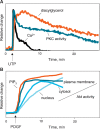
Initial activation of PKC and Akt is driven by second messenger levels but only PKC requires sustained increase in second messenger levels for sustained activity. A: Graph showing the agonist-evoked rise in intracellular Ca2+ (black line), diacylglycerol levels at the plasma membrane (orange line), and PKC activity at the plasma membrane (blue line) simultaneously reported by probes for Ca2+, diacylglycerol, and PKC activity. Initial activation is driven by Ca2+, which recruits conventional PKC to the plasma membrane, but sustained activity depends on diacylglycerol. Adapted from Gallegos et al. (33). B: Graph showing agonist-evoked rise in PIP3 levels at the plasma membrane (orange line), Akt activity at the plasma membrane (dark blue line), in the cytosol (medium blue line), and in the nucleus (light blue line). Adapted from Kunkel et al. (36).

Similar articles

Cited by

References

    1. Hurley J. H. 2006. Membrane binding domains. Biochim. Biophys. Acta. 1761 805–811. - PMC - PubMed
    1. Lemmon M. A. 2008. Membrane recognition by phospholipid-binding domains. Nat. Rev. Mol. Cell Biol. 9 99–111. - PubMed
    1. Hurley J. H., A. C. Newton, P. J. Parker, P. M. Blumberg, and Y. Nishizuka. 1997. Taxonomy and function of C1 protein kinase C homology domains. Protein Sci. 6 477–480. - PMC - PubMed
    1. Misra S., G. J. Miller, and J. H. Hurley. 2001. Recognizing phosphatidylinositol 3-phosphate. Cell. 107 559–562. - PubMed
    1. Hurley J. H., and T. Meyer. 2001. Subcellular targeting by membrane lipids. Curr. Opin. Cell Biol. 13 146–152. - PubMed

Publication types

Substances

LinkOut - more resources