. 2009 Jan;37(1):172-83.
doi: 10.1093/nar/gkn919. Epub 2008 Nov 25.

Metastases suppressor NM23-H2 interaction with G-quadruplex DNA within c-MYC promoter nuclease hypersensitive element induces c-MYC expression

Affiliations

Metastases suppressor NM23-H2 interaction with G-quadruplex DNA within c-MYC promoter nuclease hypersensitive element induces c-MYC expression

Ram Krishna Thakur et al. Nucleic Acids Res. 2009 Jan.

Abstract

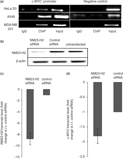
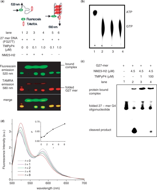
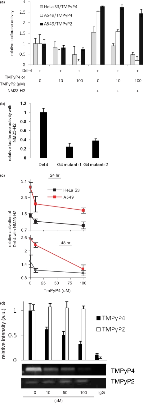
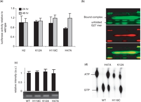
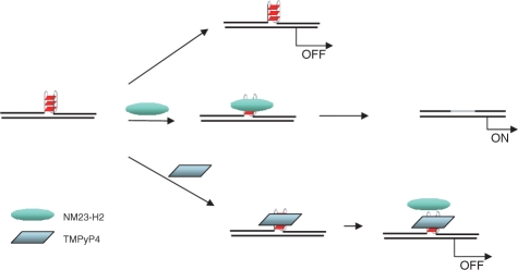
Regulatory influence of the G-quadruplex or G4 motif present within the nuclease hypersensitive element (NHE) in the promoter of c-MYC has been noted. On the other hand, association of NM23-H2 to the NHE leads to c-MYC activation. Therefore, NM23-H2 interaction with the G4 motif within the c-MYC NHE presents an interesting mechanistic possibility. Herein, using luciferase reporter assay and chromatin immunoprecipitation we show NM23-H2 mediated c-MYC activation involves NM23-H2-G4 motif binding within the c-MYC NHE. G4 motif complex formation with recombinant NM23-H2 was independently confirmed using fluorescence energy transfer, which also indicated that the G4 motif was resolved to an unfolded state within the protein-bound complex. Taken together, this supports transcriptional role of NM23-H2 via a G4 motif.


Figures

Figure 1.
Figure 1.
NM23-H2 binds c-MYC promoter in vivo (a) ChIP assays using antibodies against NM23-H2 in multiple cell lines. Immunoprecipitated DNA samples were PCR amplified to show NM23-H2 occupancy of c-MYC promoter and negative control locus. Primer and locus information are given with ChIP methods; PCR cycles were maintained in the exponential phase of amplification. (b) Semi-quantitative RT-PCR analysis of NM23-H2 mRNA expression in A549 cells treated with siRNA duplex against NM23-H2 compared to scrambled (or control) siRNA and untransfected samples. β-actin mRNA levels remain unchanged. (c and d) Quantitative real-time PCR analysis. NM23-H2 (c) and c-MYC (d) transcript levels (fold change is relative to respective transcript levels determined in cells transfected with control siRNA) in A549 cells treated with NM23-H2 siRNA or control siRNA duplex. Experiments were performed in triplicate to analyze standard deviations in all cases.
Figure 2.
Figure 2.
NM23-H2 binds to G4 motif in vitro. (a) Electrophoretic mobility shift of 27-mer oligonucleotide (FG27T, see Methods) from c-MYC promoter NHE in presence or absence of NM23-H2 and/or TMPyP4 in 5% native PAGE; protein bands were independently stained by Coomassie (data not shown). Top panel: schematic representation of energy transfer in unfolded and folded (parallel G4 motif) along with respective predominant emission wavelengths. (b) Protein function (kinase) activity of NM23-H2 was not affected in presence of TMPyP4. NM23-H2-mediated phosphorylation of GTP using [γ-32P]ATP was assayed in presence of TMPyP4. Lane 1: [γ-32P]ATP alone; lane 2: with NM23-H2; lanes 3 and 4: with NM23-H2 and 60 μM (lane 3) or 120 μM (lane 4) TMPyP4. (c) Nuclease activity of NM23-H2 is inhibited in presence of TMPyP4. Ten-nanomolar radiolabeled (along with 0.1 μM unlabeled) 27-mer oligonucleotide sequence (5′-TGG GGAGGGTGGGGAGGGTGG GGAAGG-3′) that forms G4 motif (23) in the c-MYC promoter NHE was incubated with 4.5 μM NM23-H2, either alone (lane 2), or with 1 μM (lane 3) or 100 μM (lane 4) TMPyP4 in presence of NM23-H2 in buffer [10 mM HEPES, 2 mM MgCl2, 20 mM KCl, 50 µg/ml BSA, 0.5 mM DTT (pH 7.4)]; lane 1: oligonucleotide only as control. All reactions in (b) and (c) had 60 μM single-strand poly(dA) as nonspecific control and TMPyP4 treatments were for 45 min followed by 3 h incubation with protein at 37°C. (d) FRET (detected in solution) of FG27T (50 nM) as a function of increasing concentration of NM23-H2 (r = NM23-H2/FG27T) in 20 mM K+; spectra were recorded following a 10-min incubation after each addition. Inset: Change in energy transfer (represented as P, determined from experimental FRET data) as a function of increasing (r); P = ID/(ID+IA), where ID and IA are average donor (fluorescein) and acceptor (TAMRA) intensities, respectively. Experiments were done in triplicate and for each value of P error of ± 0.07 arbitrary units was observed.
Figure 3.
Figure 3.
G4 motif is required for NM23-H2-mediated transactivation of c-MYC. (a) NM23-H2-mediated transactivation of c-MYC is inhibited in presence of TMPyP4, but not TMPyP2. Cells were transfected with Del-4 (reporter plasmid with c-MYC promoter) and treated with TMPyP4, TMPyP2, NM23-H2 expressing pCDNA3 plasmid or in combination as indicated (TMPyP4/TMPyP2 treatment was for 3 h, prior to transfection). Luciferase activity is after 48 h, normalized to Del-4 transfected untreated cells (not treated with TMPyP4/TMPyP2). (b) Disruption of G4 motif leads to decreased transactivation. NM23-H2-induced promoter activity of the reporter construct Del-4 and G4-mutant constructs (where specific substitutions that destabilize G4 motif formation were introduced within Del-4); luciferase activity is relative to NM23-H2 untransfected cells in each case. (c) Loss in transactivation in presence of TMPyP4, normalized to cells treated with same concentration of TMPyP4 but without NM23-H2. (d) ChIP with NM23-H2-specific antibody is affected in presence of TMPyP4 but not TMPyP2. Dose-dependent ChIP signal of the 165-bp c-MYC promoter NHE harboring the G4 motif sequence in HeLa S3 cells when treated with TMPyP4 or TMPyP2. IgG: ChIP using mouse IgG as nonspecific negative control.
Figure 4.
Figure 4.
Effect of NM23-H2 mutants on c-MYC transactivation. (a and b) Luciferase reporter activity of the Del-4 plasmid on co-transfection with plasmids expressing NM23-H2 (labeled as H2) and respective mutants in HeLa S3 cells (a); PAGE of complex formation of FG27T with NM23-H2 mutants detected by fluorescence (b) (lanes 1 and 5: FG27T alone; lane 2: K12A; lane 3: H118C; lane 4: H47A); (c) Enrichment of the 165-bp c-MYC promoter fragment in ChIP with NM23-H2-specific antibody for NM23-H2 wild-type (WT) and each mutant. (d) Phosphotransferase assay of mutants showing decreased phosphotransfer relative to WT activity in case of H47A; activities as shown for H118C and K12A mutants were reported earlier (52,53).
Figure 5.
Figure 5.
Proposed regulatory model for NM23-H2-G4 motif interaction in c-MYC transcription activation. Repressive influence of the G4 motif (noted earlier) is transformed in to activation of c-MYC expression on NM23-H2 binding to the G4 motif, which could be due to removal of the repressor (G4 motif) and/or recruitment of ancillary transcriptional activators. TMPyP4-binding to the G4 motif inhibits NM23-H2-G4 motif interaction possibly by disrupting the G4 motif structure.

References

    1. Hatfield GW, Benham CJ. DNA topology-mediated control of global gene expression in Escherichia coli. Annu. Rev. Genet. 2002;36:175–203. - PubMed
    1. Bacolla A, Wells RD. Non-B DNA conformations, genomic rearrangements, and human disease. J. Biol. Chem. 2004;279:47411–47414. - PubMed
    1. Rich A, Zhang S. Timeline: Z-DNA: the long road to biological function. Nat. Rev. Genet. 2003;4:566–572. - PubMed
    1. Sen D, Gilbert W. Formation of parallel four-stranded complexes by guanine-rich motifs in DNA and its implications for meiosis. Nature. 1988;334:364–366. - PubMed
    1. Balagurumoorthy P, Brahmachari SK. Structure and stability of human telomeric sequence. J. Biol. Chem. 1994;269:21858–21869. - PubMed

Publication types