. 2009 Jan;37(1):144-57.
doi: 10.1093/nar/gkn900. Epub 2008 Nov 25.

The class E floral homeotic protein SEPALLATA3 is sufficient to loop DNA in 'floral quartet'-like complexes in vitro

Affiliations

The class E floral homeotic protein SEPALLATA3 is sufficient to loop DNA in 'floral quartet'-like complexes in vitro

Rainer Melzer et al. Nucleic Acids Res. 2009 Jan.

Abstract

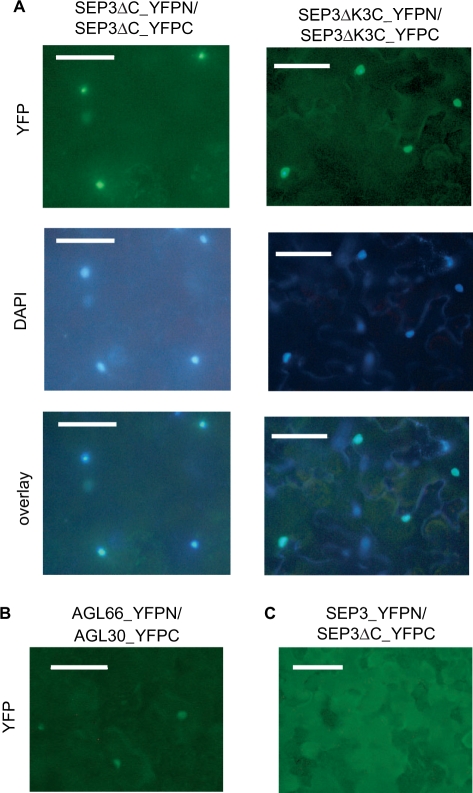
The organs of a eudicot flower are specified by four functional classes, termed class A, B, C and E, of MADS domain transcription factors. The combinatorial formation of tetrameric complexes, so called 'floral quartets', between these classes is widely believed to represent the molecular basis of floral organ identity specification. As constituents of all complexes, the class E floral homeotic proteins are thought to be of critical relevance for the formation of floral quartets. However, experimental support for tetrameric complex formation remains scarce. Here we provide physico-chemical evidence that in vitro homotetramers of the class E floral homeotic protein SEPALLATA3 from Arabidopsis thaliana bind cooperatively to two sequence elements termed 'CArG boxes' in a phase-dependent manner involving DNA looping. We further show that the N-terminal part of SEPALLATA3 lacking K3, a subdomain of the protein-protein interactions mediating K domain, and the C-terminal domain, is sufficient for protein dimerization, but not for tetramer formation and cooperative DNA binding. We hypothesize that the capacity of class E MADS domain proteins to form tetrameric complexes contributes significantly to the formation of floral quartets. Our findings further suggest that the spacing and phasing of CArG boxes are important parameters in the molecular mechanism by which floral homeotic proteins achieve target gene specificity.


Figures

Figure 1.
Figure 1.
Stoichiometry of SEP3 protein–DNA assembly. (A) EMSA in which full length (‘SEP3’) and C-terminal deleted SEP3 (‘SEP3ΔC’) were co-incubated at different ratios obtained by mixing plasmid templates in ratios of 0:1, 1:5, 1:3, 1:1, 3:1, 5:1 or 1:0. Per reaction, 2 µl of in vitro translated protein was used. DNA fragments carried either one or two CArG boxes (orange bars) as depicted at the bottom of the gel. Proteins applied are noted above the gel. AP3, that alone is not expected to bind to DNA, was used as a negative control. In lanes where no free DNA (marked by ‘0’) is visible, proteins were labelled instead of DNA for the sake of band resolution. Signals obtained with radioactively labelled DNA are shown on the right and on the left for comparison. Bands are marked with numbers (‘0’, ‘2’ and ‘4’) according to the number of proteins bound to the DNA fragment; lowercase letters are used to differentiate between complexes composed of different proteins. The inferred complex composition is shown on the right. Full length proteins are shown in green, truncated ones in yellow. ‘M’ denotes marker lanes in which a radioactively labelled DNA ladder (100-bp DNA ladder, NEB) was applied. All signals were obtained from a single gel, but exposure time for lanes containing radioactively labelled DNA fragments was different from the rest. (B) Proposed mechanism of MADS domain protein–DNA assembly. Binding of the first protein dimer to a CArG box is characterized by the dissociation constant Kd1, binding of the second dimer is characterized by the dissociation constant Kd2. Binding of the second dimer can be independent of binding of the first dimer, or cooperative and involving DNA looping.
Figure 2.
Figure 2.
Analysis of cooperative DNA binding of SEP3. (A and B) Examples of EMSAs used to determine cooperative DNA binding of SEP3. Different protein concentrations were added to a DNA probe carrying two CArG boxes. (A) A DNA probe carrying two CArG boxes spaced by 6 helical turns was used. (B) Spacing between the CArG boxes was 6.5 helical turns. Comparing the gel pictures shown in (A) and (B), the difference in cooperative binding is evident by the stronger signal caused by a DNA-bound SEP3 dimer (‘2’) in (B). (DNA fragments to which a single SEP3 dimer is bound migrate more slowly at increased protein concentration possibly due to higher glycerol concentrations in these samples.) For size comparison, a SEP3 dimer bound to a probe containing only one CArG box is shown always on the left. Quantitative analysis showing the fractional saturation of the different bands (circles: free DNA; triangles: one dimer bound; squares: two dimer/tetramer bound) is shown below each gel picture. Binding curves were calculated as described in Materials and methods section. (C) Phase dependence of the formation of the SEP3–DNA complex. The binding cocktail contained 0.4 µl of in vitro translated protein. Signals resulting from complexes bound to probes containing one CArG box are shown in the leftmost lanes. A dimer bound to a CArG box peripheral located on the DNA fragment is indicated by ‘2’, whereas ‘2′’ indicates a dimer bound to a CArG box centrally located on the DNA fragment. The difference in migration of the two complexes results in part from the ability of SEP3 to bend DNA. Below the gel picture, quantitative analysis of homotetramer formation is shown. Fractional saturation of the signal intensity caused by the homotetramer is expressed as percentage of the fractional saturation of the homotetramer bound to the CArG boxes spaced by 6 helical turns. Band assignment is as in Figure 1.
Figure 3.
Figure 3.
DNase I footprint assays. SEP3 or SEP3ΔK3C was incubated with a DNA probe carrying two CArG boxes spaced by 6 helical turns. For SEP3ΔK3C, but not for SEP3, a complex with one dimer bound could be obtained at sufficient signal intensity and thus was also analysed on the sequencing gel. Protein–DNA complexes analysed are noted above the gel. An A+G sequencing reaction of the DNA probe used is shown for comparison. Sequence of the DNA is depicted on the right (blue = cytosine, red = thymine, green = adenine, black = guanine); the position of the CArG boxes is indicated. Open and filled arrowheads point towards sites of diminished and enhanced DNase I sensitivity, respectively. On the right, quantitative analysis of the gel picture shows the change of sensitivity to DNase I digestion after protein binding, in single base pair steps, using free DNA as a reference after correction for differences in DNA-loading by using invariable bands as an internal standard. The region protected by full-length SEP3 extends beyond the CArG boxes, probably because these DNA regions are in close contact to the protein surface in the looped complex.
Figure 4.
Figure 4.
Examples of EMSAs used to determine cooperativity of DNA binding of SEP3ΔC and SEP3ΔK3C. (A–D) show EMSAs in which increasing amounts of SEP3ΔC (A and C) or SEP3ΔK3C (B and D) were incubated with DNA probes carrying two CArG-boxes spaced by 6 (A and B) or 6.5 (C and D) helical turns. As in Figure 2, quantitative analysis of the band pattern is shown below each gel picture. The ability of the proteins to bind as dimers to a DNA fragment carrying one CArG box is shown in the leftmost lanes of (A) and (B). (E) Analysis of the phasing of two CArG boxes on the formation of a SEP3ΔC–DNA and SEP3ΔK3C–DNA complex. 0.4 μl of in vitro translated protein was used. Signals resulting from complexes bound to probes containing one CArG box are shown in the leftmost lanes. A dimer bound to a CArG box peripheral located on the DNA fragment is indicated by ‘2’, whereas ‘2′’ indicates a dimer bound to a CArG box centrally located on the DNA fragment. As both, SEP3ΔC and SEP3ΔK3C, bend DNA, the differential positioning of the CArG box on the fragment results in a difference in migration of the complexes (2 and 2′). As in Figure 2C, quantitative analysis of the complex consisting of four proteins bound to DNA is shown below the gel picture.
Figure 5.
Figure 5.
(A) Subcellular localization and in planta dimerization of SEP3ΔC and SEP3ΔK3C. Proteins are expressed as fusions with the N- (YFPN) or C-terminal (YFPC) part of YFP as indicated above the picture in Nicotiana benthamiana leaves. Leaf sections were analysed with a fluorescence microscope. YFP signals are shown in the upper row, nuclear staining using DAPI is shown in the middle row. The overlay in the lower row shows co-localization of the DAPI and YFP signals. (B) The YFP signals of two proteins known to interact, i.e. AGL30 (fused to the C-terminal part of YFP) and AGL66 (fused to the N-terminal part of YFP) is shown as a positive control. (C) No YFP signal could reliably be detected when full-length SEP3 was tested alone or in combination with a C-terminal deleted version, as exemplarily shown for SEP3 (fused to the N-terminal part of YFP) co-expressed with SEP3ΔC (fused to the C-terminal part of YFP). This might be due to steric hindrance of the C-terminal domain. Scale bars, 100 µm.
Figure 6.
Figure 6.
Circular permutation analysis of DNA distortions induced by SEP3 and SEP3ΔK3C. The gel picture shows EMSAs with SEP3 and SEP3ΔK3C bound to circularly permutated probes. In vitro translated SEP3 and SEP3ΔK3C of 4 µl and 2 µl, respectively, were used. DNA probes and restriction endonucleases used to prepare them are schematically depicted above the gel. The CArG box is indicated by an orange bar. In the diagram below the gel, the complex mobility is plotted against the relative position of the CArG boxes. Bending angles represent the means of at least four different experiments (standard error in brackets). Band assignment is as in Figure 1.

References

    1. Hughes CL, Kaufman TC. Hox genes and the evolution of the arthropod body plan. Evol. Dev. 2002;4:459–499. - PubMed
    1. Veraksa A, Del Campo M, McGinnis W. Developmental patterning genes and their conserved functions: from model organisms to humans. Mol. Genet. Metabol. 2000;69:85–100. - PubMed
    1. Haughn GW, Somerville CR. Genetic control of morphogenesis in Arabidopsis. Dev. Genet. 1988;9:73–89.
    1. Coen ES, Meyerowitz EM. The war of the whorls - genetic interactions controlling flower development. Nature. 1991;353:31–37. - PubMed
    1. Mandel MA, Gustafson-Brown C, Savidge B, Yanofsky MF. Molecular characterization of the Arabidopsis floral homeotic gene APETALA1. Nature. 1992;360:273–277. - PubMed

Publication types