. 2009 Jun;11(3):250-9.
doi: 10.1215/15228517-2008-095. Epub 2008 Nov 25.

Antitumor effect in medulloblastoma cells by gefitinib: Ectopic HER2 overexpression enhances gefitinib effects in vivo

Affiliations

Antitumor effect in medulloblastoma cells by gefitinib: Ectopic HER2 overexpression enhances gefitinib effects in vivo

Daniela Meco et al. Neuro Oncol. 2009 Jun.

Abstract

The effects of the epidermal growth factor receptor (EGFR) inhibitor gefitinib on cell growth and signaling were evaluated in three medulloblastoma (MB) cell lines (D283, D341, Daoy), one supratentorial primitive neuroectodermal tumor cell line (PFSK), and four MB primary cultures. Cell lines showed diverse expression of EGFR and human epidermal receptor 2 (HER2), with high levels of constitutively activated HER2 in the HER2-overexpressing D341 and D283 cells. Gefitinib sensitivity varied across lines and was not related to expression of HER receptors or receptor baseline activation. Gefitinib induced G(0)/G(1) arrest in all lines, whereas apoptosis was dose-dependently induced only in D283 and D341 cells. The molecular response to gefitinib was investigated in Daoy and D341 lines, which showed a higher (half-maximal inhibitory concentration [IC(50)], 3.8 microM) and lower (IC(50), 6.6 microM) sensitivity to the agent, respectively. Gefitinib inhibited constitutive and EGF-triggered EGFR phosphorylation in both lines but was ineffective in constitutive activation of HER2 in D341 cells. Phosphorylated AKT inhibition paralleled that of phosphorylated EGFR, suggesting the presence of an autocrine gefitinib-sensitive EGFR/AKT pathway. On the whole, EGF-dependent signaling was less responsive to ligand stimulation and gefitinib inhibition in D341 cells, which correlated with the lower sensitivity to gefitinib's antiproliferative effect of this line. In vivo, the growth of D341 and Daoy xenografts treated with gefitinib at 150 mg/kg per day was inhibited by approximately 50%. Ectopically overexpressed HER2 in Daoy cells significantly increased sensitivity to gefitinib's antitumor effects in vivo (tumor volume inhibition = 78%). Our data indicate that gefitinib might be a molecularly targeted agent for the treatment of MB.


Figures

Fig. 1
Fig. 1
Time- and dose-dependent inhibition of the growth of medulloblastoma (MB) cell lines by gefitinib. D283 (A), D341 (B), PFSK (C), and Daoy (D) cells were exposed to the indicated concentrations of gefitinib (μM) for up to 5 days. Viable cells were counted daily. Each point represents the average value of two independent experiments, each in triplicate. Dashed lines indicate the 50% growth inhibition.
Fig. 2
Fig. 2
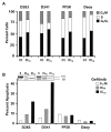
(A) Effects of gefitinib on cell cycle distribution in medulloblastoma cell lines. Cells were treated with gefitinib at equitoxic doses (Inhibitory concentration 70 [IC70] values) for 48 h, and cell cycles were assessed by flow cytometry. The percentages of the total cell population in the different phases of the cell cycle are indicated. Ct, control. (B) Apoptosis assay. Cells were treated with IC50 and IC70 concentrations of gefitinib and harvested at 72 h. Apoptosis was measured as sub-G1 content by flow cytometry. The figure shows one representative experiment in duplicate. Inset: Western blot analysis of poly(ADP-ribose) polymerase (PARP) cleavage in D283 and D341 cells treated with gefitinib in a parallel experiment.
Fig. 3
Fig. 3
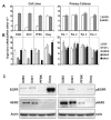
Expression of receptors and ligands of the HER family in medulloblastoma (MB) cell lines. (A) Real-time quantitative reverse transcriptase (RT)-PCR analysis of expression of epidermal growth factor receptor (EGFR) and HER2. (B) Real-time quantitative RT-PCR analysis of expression of ligands EGF, transforming growth factor α (TGFα), amphiregulin (AREG), epiregulin (EREG), and neuregulin 1 (NRG1) in MB cell lines and primary cultures. Bars show means + SD of three determinations of each target gene normalized to the endogenous control GAPDH in each sample. (C) Expression levels of total and activated EGFR and HER2 in MB cell lines. Cells were serum-starved overnight in 0.5% fetal bovine serum medium, and Western blot analysis was performed on either the phosphorylated or total levels of each protein. Equal loading was verified by actin immunoblotting.
Fig. 4
Fig. 4
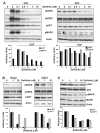
Dose-dependent inhibition of mitogen-activated protein kinase (MAPK) and AKT kinase pathways by gefitinib in Daoy and D341 cell lines. (A) Serum-starved cells were incubated with the indicated concentrations of gefitinib and then stimulated with epidermal growth factor (EGF) for 30 min. Membranes were immunostained for phosphorylated EGFR (pEGFR), pHER2, pMAPK, and pAKT. Actin was used as a loading control. (B) Effects of gefitinib on baseline EGFR, HER2, and AKT phosphorylation in Daoy and D341 cells. Serum-starved cells were incubated with the indicated concentrations of gefitinib for 2 h. (C) Dose-dependent inhibition by gefitinib of AKT and MAPK phosphorylation in Daoy cells treated with 1, 5, and 10 μM gefitinib for 24 h in 10% fetal bovine serum medium. Relative changes of the phosphorylation of AKT or MAPK (A), EGFR and AKT (B), and AKT and MAPK (C) were quantified by densitometric analysis and normalized to the densitometric value of actin. One representative experiment is shown.
Fig. 5
Fig. 5
Antitumor activity of gefitinib on human medulloblastoma xenografts. (A and B) Mice bearing Daoy (A) or D341 (B) xenografts received either vehicle only or daily oral gefitinib at 150 mg/kg per day for 5 consecutive days for 4 consecutive weeks. The treatment was started 11 and 7 days following cell implantation. Points shown are mean ± SE values for groups of 8–10 mice. TVI, tumor volume inhibition. Tumor volume was determined twice per week. (C and D) Effects of gefitinib treatment on the body weight changes of animals bearing Daoy (C) and D341 (D) tumors.
Fig. 6
Fig. 6
Ectopically overexpressed HER2 increases the sensitivity to gefitinib of Daoy xenografts. (A) Western blot analysis of total HER2 and pHER2 in parental Daoy cells and cells transfected with empty vector or pcDNA3.1/HER2 (clones 28 and 36). (B) Antitumor activity of gefitinib in Daoy/HER2 xenografts. Mice were orally administered gefitinib as described in Fig. 5. Points shown are mean ± SE values for groups of 8–10 mice. (C) Western blot analysis of EGFR, HER2, and pHER2 expression in Daoy/vector and Daoy/HER2 xenografts in animals treated with vehicle or oral gefitinib. (D) Histological analysis of tumor Daoy/HER2 xenograft tissue harvested from mice treated with vehicle or gefitinib shows a marked loss of neoplastic cells and abundant multinucleated foamy macrophages.

References

    1. Wechsler-Reya R, Scott MP. The developmental biology of brain tumors. Annu Rev Neurosci. 2001;24:385–428. - PubMed
    1. Gilbertson RJ. Medulloblastoma: Signalling a change in treatment. Lancet Oncol. 2004;5:209–218. - PubMed
    1. Luttjeboer M, Kaspers GJ. Medulloblastoma: Need for targeted treatment. Expert Rev Anticancer Ther. 2006;6:649–652. - PubMed
    1. Olayioye MA, Neve RM, Lane HA, Hynes NE. The ErbB signaling network: Receptor heterodimerization in development and cancer. EMBO J. 2000;19:3159–3167. - PMC - PubMed
    1. Yarden Y, Sliwkowski MX. Untangling the ErbB signalling network. Nat Rev Mol Cell Biol. 2001;2:127–137. - PubMed

Publication types

MeSH terms