. 2008 Dec 9;105(49):19300-5.
doi: 10.1073/pnas.0803992105. Epub 2008 Nov 25.

MicroRNA-184 antagonizes microRNA-205 to maintain SHIP2 levels in epithelia

Affiliations

MicroRNA-184 antagonizes microRNA-205 to maintain SHIP2 levels in epithelia

Jia Yu et al. Proc Natl Acad Sci U S A. .

Abstract

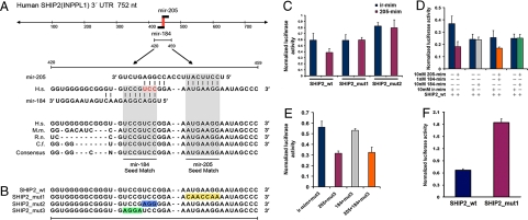
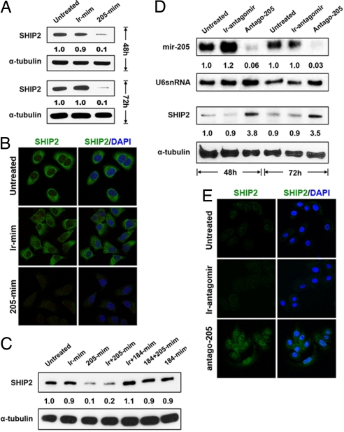
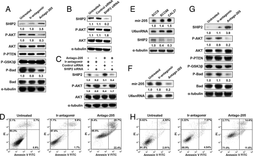
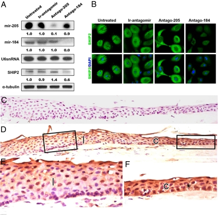
Despite their potential to regulate approximately one-third of the whole genome, relatively few microRNA (miRNA) targets have been experimentally validated, particularly in stratified squamous epithelia. Here we demonstrate not only that the lipid phosphatase SHIP2 is a target of miRNA-205 (miR-205) in epithelial cells, but, more importantly, that the corneal epithelial-specific miR-184 can interfere with the ability of miR-205 to suppress SHIP2 levels. This is the first example of a miRNA negatively regulating another to maintain levels of a target protein. Interfering with miR-205 function by using a synthetic antagomir, or by the ectopic expression of miR-184, leads to a coordinated damping of the Akt signaling pathway via SHIP2 induction. This was associated with a marked increase in keratinocyte apoptosis and cell death. Aggressive squamous cell carcinoma (SCC) cells exhibited elevated levels of miR-205. This was associated with a concomitant reduction in SHIP2 levels. Partial knockdown of endogenous miR-205 in SCCs markedly decreased phosphorylated Akt and phosphorylated BAD levels and increased apoptosis. We were able to increase SHIP2 levels in SCC cells after inhibition of miR-205. Therefore, miR-205 might have diagnostic value in determining the aggressivity of SCCs. Blockage of miR-205 activity with an antagomir or via ectopic expression of miR-184 could be novel therapeutic approaches for treating aggressive SCCs.


Conflict of interest statement

The authors declare no conflict of interest.

Figures

Fig. 1.
Fig. 1.
miR-205 targets SHIP2 at 3′ UTR and can be regulated by miR-184. (A) Sequence of the miR-205 and miR-184 binding sites within the human SHIP2 (INPPL1) 3′ UTR. Red nucleotides are overlapping binding sites. Shaded areas represent conserved complementary nucleotides of miR-184 and miR-205 seed sequences in various mammals (H.s, human; M.m, mouse; R.n, rat; C.f, chicken). (B) Schematic of the reporter constructs showing entire 3′ UTR SHIP2 sequence (SHIP2 _wt) and the mutated 3′ UTR nucleotides (yellow) of the miR-205 binding site (SHIP2_mut1). SHIP2_mut2 represents the reporter construct containing mutated overlapping nucleotides (blue) of miR-184 and miR-205. SHIP2_mut3 represents the reporter construct containing nucleotides (green) predicted to be exclusively used for miR-184 binding to SHIP2 mRNA. (C) Luciferase activity of (i) SHIP2_wt in the presence of 10 nM of miR-205 showing the inhibitory activity of this reporter and (ii) the SHIP2_mut1 and mut2 reporters, showing that miR-205 mimic cannot inhibit the luciferase activity of these constructs compared with the wild-type construct. The overlapping nucleotides of miR-184 and miR-205 are required for miR-205 binding to SHIP2 3′ UTR as is the seed sequence of miR-205. Error bars (SEM) are derived from six experiments in triplicate. (D) Luciferase activity of SHIP2_wt reporter in the presence (+) or absence (−) of various concentrations of miR-205, miR-184, or nontargeting (irrelevant) mimics. Transfection (red) of miR-205 mimic inhibits luciferase activity, whereas transfection (gray) of miR-184 mimic has no effect. Cotransfection (orange) of 1 nM miR-184 and 10 nM miR-205 mimics cannot completely restore luciferase activity, whereas cotransfection (green) of equal amounts of miR-184 and miR-205 mimics completely rescued luciferase activity. Error bars (SEM) are derived from three experiments in triplicate. (E) Luciferase activity of SHIP2_mut3 reporter showing that (i) this mutation does not inhibit miR-205 binding to SHIP2 3′ UTR (blue and red columns); (ii) miR-184 does not inhibit this mutated reporter (gray column); and (iii) cotransfection of miR-184 and miR-205 cannot restore luciferase activity of 184 mut3 (orange column). Error bars (SEM) are derived from three experiments in triplicate. Controls for these experiments are shown in Fig. S1 C and D. (F) Luciferase activity of SHIP2_wt and SHIP2_mut1 in HEKs showing that endogenous miR-205 inhibits SHIP2. Positive controls (184/205_PER) are shown in Fig. S1E.
Fig. 2.
Fig. 2.
SHIP2 levels are controlled by miR-205 and miR-184. (A) Immunoblotting of SHIP2 in HeLa cells that were treated with a miR-205 mimic, decrease protein 48 and 72 h after treatment. (B) Immunofluorescence microscopy of HeLa cells stained with anti-SHIP2 and anti-SHIP2/DAPI showing a marked decrease in staining 72 h after treatment with miR-205 mimic. Staining data at 48 h is presented in Fig. S2A. (C) Immunoblotting of SHIP2 in HeLa cells that were untreated (1), transfected with an irrelevant mimic (ir-mim; 2), miR-205 mimic (205-mim; 3), miR-205 mimic plus and irrelevant mimic (ir + 205-mim; 4), miR-184 mimic plus an irrelevant mimic (ir + 184-mim; 5), miR-184 plus miR-205 mimics (184 + 205-mim;6), and miR-184 mimic (184-mim; 7) for 48 h. miR-205 mimic reduces SHIP2 levels (3, 4) whereas miR-184 inhibits miR-205 from reducing SHIP2 levels (6). (D) Northern analysis using a miR-205 specific probe showing a marked decrease in miR-205 levels in HEKs treated with an antagomir to miR-205 (Antago-205) for 48 and 72 h. Immunoblotting of SHIP2 and α-tubulin in HEKs shows an increase in SHIP2 expression 48 and 72 h after treatment with Antago-205. (E) Immunofluorescence microscopy of HEKs stained with SHIP2 showing an increase in staining after 72 h of treatment with Antago-205. Staining data at 48 h are presented in Fig. S2B. Numbers below the panels represent the normalized expression signal of proteins and RNAs.
Fig. 3.
Fig. 3.
miR-205 affects the Akt pathway in keratinocytes directly through targeting of SHIP2 and is inversely correlated with SHIP2 in SCC cell lines. (A) Immunoblotting of SHIP2, phosphorylated Akt (p-Akt), total pan (1/2/5) Akt, phosphorylated BAD, total BAD, phosphorylated PTEN (p-PTEN), and phosphorylated GSK3β (p-GSK3β) in HEKs that were untreated (un-rx) or treated with an ir-antagomir or Antago-205 for 48 h. α-Tubulin serves as a loading control. (B) Immunoblots of SHIP2, p-Akt, AKT, and α-tubulin in HEKs 72 h after transfection with SHIP2 siRNA and control siRNA, showing decreases in SHIP2 and increases in p-Akt. (C) Immunoblots of SHIP2, p-Akt, AKT, and α-tubulin in HEKs 48 h after treatment with an antagomir to miR-205 or an irrelevant antagomir. HEKs were subsequently treated for another 72 h with combinations of siRNA to SHIP2, control siRNA, antagomir-205, and irrelevant antagomir. (D) Keratinocytes were stained with propidium iodide and annexin V 48 h after treatment with an ir-antagomir or Antago-205 and compared with untreated cells. Late apoptotic cells are seen in the top right quandrant. (E) Northern analysis of oral SCC cell lines using a miR-205-specific probe showing increases in miR-205 in SCC68 and CAL27 cells. Immunoblotting of SHIP2 in oral SCCs showing a marked decrease in SHIP2 in SCC68 and CAL27 cells. (F) Northern analysis with a miR-205-specific probe in SCC68 cells that were treated with an ir-antagomir or Antago-205 for 48 h. U6 serves as a loading control. (G) Immunoblotting of SHIP2, p-Akt, total Akt, p-PTEN, p-GSK3β, p-BAD, and BAD in SCC68 cells treated as described in F. α-Tubulin serves as loading control. (H) SCC68 cells were treated as described in F and G and then stained with propidium iodide and annexin V. Numbers below the panels represent the normalized expression signal of proteins and RNAs.
Fig. 4.
Fig. 4.
miR-184 alters the ability of miR-205 to affect SHIP2 in corneal keratinocytes in vitro and in vivo. (A) Northern analysis of primary human corneal epithelial (HCEKs) cells using specific probes for miR-184 and miR-205, showing expression of both of these miRNAs in untreated and control (Ir-antagomir) cells. After 72 h of treatment, each antagomir abrogates the respective miRNA. U6 serves as a loading control. Shown is immunoblotting of SHIP2 and α-tubulin in HCEKs that were untreated or were treated with Ir-antagomir, Antago-205, or an antagomir to miR-184 (Antago-184) for 72 h. (B) Immunofluorescence microscopy of HCEKs stained for SHIP2 showing a marked decrease in staining after a 72-h treatment with antagomir-184, whereas treatment with antagomir to miR-205 resulted in an increase in SHIP2 staining. (C and D) Serial frozen sections of human limbal and corneal epithelium immunohistochemically stained with an antibody that recognizes IgG (control, C) or SHIP2 (D). (E and F) Higher magnification of the boxed areas of the limbal (l, E) and corneal (c, F) epithelia, showing a decrease in SHIP2 staining in the limbal epithelium compared with the corneal epithelium. Numbers below the panels represent the normalized expression signal of proteins and RNAs.
Fig. 5.
Fig. 5.
Proposed regulatory effects of miR-205 and miR-184 on SHIP2 levels in various epithelial contexts. (A) Epidermal keratinocytes. Decreasing miR-205 via antagomir-205 increases SHIP2 levels resulting in the dampening of Akt signaling and an increase in apoptosis and cell death. (B) Corneal keratinocytes. Decreasing miR-184 via antagomir-184 “releases” miR-205 to reduce SHIP2 levels augmenting the Akt pathway, with increased cell survival and angiogenesis as possible outcomes. Because miR-184 does not inhibit SHIP2, decreasing miR-205 via antagomir-205 disturbs the normal balance between SHIP2 and miR-184/205. (C) SCC. Ectopic expression of miR-184 or treatment with an antagomir to miR-205 represents potential therapeutic modalities for the treatment of SCCs by increasing SHIP2 levels, which might act as a tumor suppressor in these neoplasias.

References

    1. Ambros V. The functions of animal microRNAs. Nature. 2004;431:350–355. - PubMed
    1. Lee RC, Feinbaum RL, Ambros V. The C. elegans heterochronic gene lin-4 encodes small RNAs with antisense complementarity to lin-14. Cell. 1993;75:843–854. - PubMed
    1. Esau C, et al. MicroRNA-143 regulates adipocyte differentiation. J Biol Chem. 2004;279:52361–52365. - PubMed
    1. Hornstein E, et al. The microRNA miR-196 acts upstream of Hoxb8 and Shh in limb development. Nature. 2005;438:671–674. - PubMed
    1. Sempere LF, et al. Expression profiling of mammalian microRNAs uncovers a subset of brain-expressed microRNAs with possible roles in murine and human neuronal differentiation. Genome Biol. 2004;5:R13.11–R13.11. - PMC - PubMed

Publication types

MeSH terms

Substances