. 2008 Dec 16;105(50):19726-31.
doi: 10.1073/pnas.0803488105. Epub 2008 Nov 25.

Electron tomography of early melanosomes: implications for melanogenesis and the generation of fibrillar amyloid sheets

Affiliations

Electron tomography of early melanosomes: implications for melanogenesis and the generation of fibrillar amyloid sheets

Ilse Hurbain et al. Proc Natl Acad Sci U S A. .

Abstract

Melanosomes are lysosome-related organelles (LROs) in which melanins are synthesized and stored. Early stage melanosomes are characterized morphologically by intralumenal fibrils upon which melanins are deposited in later stages. The integral membrane protein Pmel17 is a component of the fibrils, can nucleate fibril formation in the absence of other pigment cell-specific proteins, and forms amyloid-like fibrils in vitro. Before fibril formation Pmel17 traffics through multivesicular endosomal compartments, but how these compartments participate in downstream events leading to fibril formation is not fully known. By using high-pressure freezing of MNT-1 melanoma cells and freeze substitution to optimize ultrastructural preservation followed by double tilt 3D electron tomography, we show that the amyloid-like fibrils begin to form in multivesicular compartments, where they radiate from the luminal side of intralumenal membrane vesicles. The fibrils in fully formed stage II premelanosomes organize into sheet-like arrays and exclude the remaining intralumenal vesicles, which are smaller and often in continuity with the limiting membrane. These observations indicate that premelanosome fibrils form in association with intralumenal endosomal membranes. We suggest that similar processes regulate amyloid formation in pathological models.


Conflict of interest statement

The authors declare no conflict of interest.

Figures

Fig. 1.
Fig. 1.
Ultrastructure of MNT-1 cells using HPF and freeze substitution. (Upper) A low-magnification image showing mature melanosomes (stages IV and III) at the periphery and immature melanosomes (stages II and I) in the Golgi area. PM, plasma membrane; GA, Golgi Apparatus; m, mitochondria; ER, endoplasmic reticulum. (Lower) Selected images of stage I, II, III, and IV melanosomes. (Scale bars: 200 nm.)
Fig. 2.
Fig. 2.
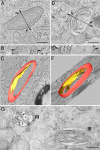
The intralumenal fibrils of stage II premelanosomes are organized as sheets. (A) Slice of an electron tomographic reconstruction of a stage II premelanosome. The line indicates the axis of rotation for visualization along the z axis in B. (A and B) The intralumenal striations follow the curvature of the melanosomal membrane (arrows), revealing a sheet-like morphology. (C) Three-dimensional model of the melanosomal membrane (in magenta) and one single fibril (in yellow) (see Movie S1). (D) Slice of a reconstruction of a stage III melanosome. The line indicates the axis of rotation for visualization along the z axis in E. (D and E) Note the melanin deposits over the sheets. (F) Three-dimensional model of the melanosomal membrane (in magenta) and one single fibril with melanin (in yellow) (see Movie S2). (G, Left) A cross section through an early stage III melanosome reveals the sheet-like organization of the melanin-coated fibrillar structures (Left, arrows) as compared to the elongated fibrillar appearance of the longitudinally sectioned melanosome (Right). (Scale bars: 200 nm.)
Fig. 3.
Fig. 3.
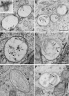
Fibrils begin to form on ILVs within early MVBs. Small fibrils (arrows) are closely associated to nascent buds (A, see Inset for higher magnification) and vesicles of early MVBs (B, C, and D). (B) Examples of MVBs in which thin fibrils are seen emanating from 60–70 nm intralumenal vesicles (arrows). (C and D) Examples of organelles bearing smaller, 40-nm vesicles (arrowheads) and more organized fibrils (arrows). (E) An example of an ellipsoidal stage II premelanosome with organized arrays of fibrils (II). (F) Note the sheet-like appearance of the fibrils lying parallel to the plane of the section (double arrows). (Scale bars: 200 nm.)
Fig. 4.
Fig. 4.
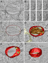
Fibrils are closely associated with ILVs of early MVBs. (A and B) A tomogram slice (A) and sequential z axis slices through the tomographic volume (B) showing close association of small fibrils (arrows) with intralumenal vesicles. (C and D) Membranes and fibrils in consecutive tomogram slices are manually contoured; one slice is shown in C, and the 3D model is shown in D. The limiting membrane of the organelle is in red, vesicles with clearly associated fibrils (yellow) are in blue and those with no obvious fibrils are in green. Closely apposed endoplasmic reticulum membranes are in white. (E) The ILVs and associated fibrils are highlighted and shown rotated. Note the short fibrils emanating from larger ILVs. Arrow indicates the vesicle shown in F. (F) Computationally detected fibrils emanating from a large ILV. See also Movie S3A for tilt series and tomographic reconstruction, Movie S3B for 3D model, and Movie S3C for representation of fibrils by isosurfacing. (Scale bar: 200 nm.)
Fig. 5.
Fig. 5.
Stage II premelanosomes bear ILVs continuous with the limiting membrane. (A and B) A tomogram slice (A) and sequential z axis slices through the tomographic volume (B) illustrating an internal vesicle fused with the limiting membrane of a stage II melanosome. (C and D) Membranes and fibrils in consecutive slices from the tomogram are manually contoured; one slice is shown in C, and the 3D model is shown in D. The limiting membrane of the organelle is in red; free vesicles are in green and those fusing with the membrane in yellow. Fibrils are in brown. Closely apposed endoplasmic reticulum membranes are in white. (E and F) The melanosomal membrane and the ILVs are highlighted. Note the continuities between the ILVs (yellow) and the melanosomal membrane, indicated by the appearance of the ILV membrane on the cytosolic face of the melanosome (arrows). See also Movie S4A for tilt series and tomographic reconstruction, Movie S4B for manual contouring, and Movie S4C for 3D model. (Scale bar: 200 nm.)

References

    1. Seiji M, Fitzpatrick TM, Simpson RT, Birbeck MSC. Chemical composition and terminology of specialized organelles (melanosomes and melanin granules) in mammalian melanocytes. Nature. 1963;197:1082–1084. - PubMed
    1. Raposo G, Tenza D, Murphy DM, Berson JF, Marks MS. Distinct protein sorting and localization to premelanosomes, melanosomes, and lysosomes in pigmented melanocytic cells. J Cell Biol. 2001;152:809–824. - PMC - PubMed
    1. Raposo G, Marks MS. Melanosomes–dark organelles enlighten endosomal membrane transport. Nat Rev Mol Cell Biol. 2007;8:786–797. - PMC - PubMed
    1. Chakraborty AK, et al. Polymerization of 5,6-dihydroxyindole-2-carboxylic acid to melanin by the pmel 17/silver locus protein. Eur J Biochem. 1996;236:180–188. - PubMed
    1. Lee ZH, et al. Characterization and subcellular localization of human Pmel 17/silver, a 100-kDa (pre)melanosomal membrane protein associated with 5,6,-dihydroxyindole-2-carboxylic acid (DHICA) converting activity. J Invest Dermatol. 1996;106:605–610. - PubMed

Publication types

MeSH terms

LinkOut - more resources