Comparative Study
. 2008 Dec 9;105(49):19514-9.
doi: 10.1073/pnas.0809662105. Epub 2008 Nov 25.

Comparing face patch systems in macaques and humans

Affiliations
Comparative Study

Comparing face patch systems in macaques and humans

Doris Y Tsao et al. Proc Natl Acad Sci U S A. .

Abstract

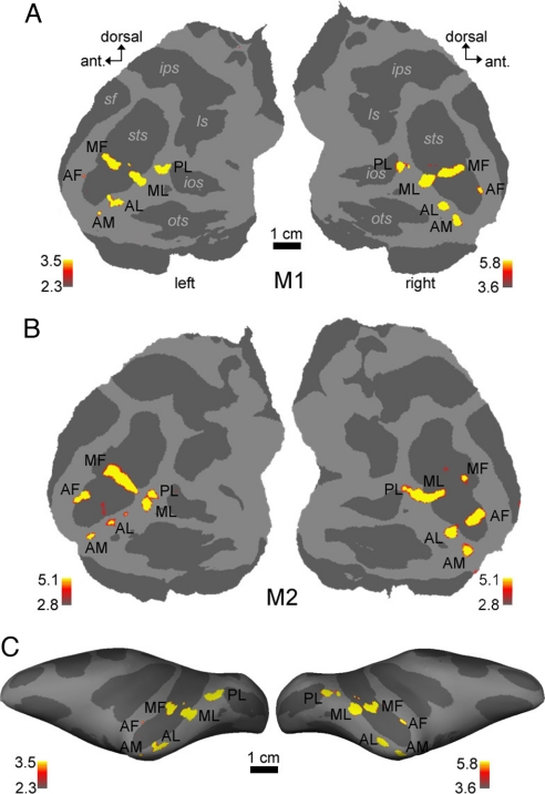
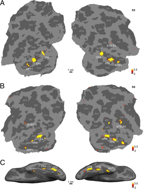
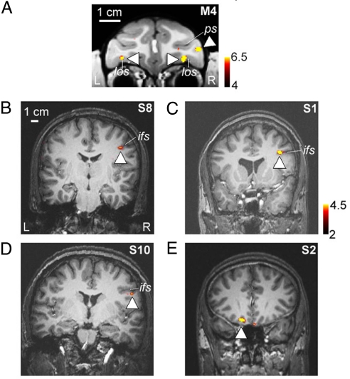
Face recognition is of central importance for primate social behavior. In both humans and macaques, the visual analysis of faces is supported by a set of specialized face areas. The precise organization of these areas and the correspondence between individual macaque and human face-selective areas are debated. Here, we examined the organization of face-selective regions across the temporal lobe in a large number of macaque and human subjects. Macaques showed 6 regions of face-selective cortex arranged in a stereotypical pattern along the temporal lobe. Human subjects showed, in addition to 3 reported face areas (the occipital, fusiform, and superior temporal sulcus face areas), a face-selective area located anterior to the fusiform face area, in the anterior collateral sulcus. These results suggest a closer anatomical correspondence between macaque and human face-processing systems than previously realized.


Conflict of interest statement

The authors declare no conflict of interest.

Figures

Fig. 1.
Fig. 1.
Face-selective regions in 2 macaques, superimposed on flattened cortical surfaces (A and B) and a lateral view of the inflated hemispheres (C). Computational flattening involves distorting the spatial arrangement of the data and underestimates the size of the sulci (shown in dark gray). Color scale bars indicate the negative common logarithm of the probability of error. PL, posterior face patch; MF, middle face patch in the STS fundus; ML, middle face patch on the STS lip; AF, anterior face patch in the STS fundus; AL, anterior face patch on the STS lip; AM, anterior face patch on the ventral surface of IT just lateral and anterior to the AMTS; ls, lunate sulcus; ios, inferior occipital sulcus; ots, occipitotemporal sulcus; sts, superior temporal sulcus; ips, intraparietal sulcus; sf, Sylvan fissure.
Fig. 2.
Fig. 2.
Face-selective regions in the left and right temporal lobes of 2 humans, superimposed on flattened cortical surfaces (A and B) and a ventral view of the inflated hemispheres (C). OFA, occipital face area; FFA, fusiform face area; STS-FA, superior temporal sulcus face area; AFP1, anterior face patch 1; aos, anterior occipital; los, lateral occipital; lots, lateral occipitotemporal; ips, intraparietal; its, inferior temporal; sts, superior temporal; pos, parietooccipital; cas, calcarine; cos, collateral; tos, transoccipital; pcs, precentral; and sf, Sylvan fissure.
Fig. 3.
Fig. 3.
Prefrontal face-selective regions in macaque and humans. (A) Prefrontal face patches from 1 macaque (M4), shown on a coronal slice (taken from ref. 51). The monkey had a face patch in the lateral orbitofrontal sulcus bilaterally, and 1 face patch in the inferior convexity, within the infraprincipal dimple, exclusively in the right hemisphere. los, lateral orbitofrontal sulcus; ps, principal sulcus. (B–D) Lateral prefrontal face-selective regions from 3 human subjects (S8, S1, and S10), shown on coronal slices. The location of the human lateral prefrontal face-selective region was consistent across subjects (within the inferior frontal sulcus, strongly lateralized to the right hemisphere). (E) Face-selective region in orbitofrontal cortex of a human subject (S2).

References

    1. Darwin C. The Expression of the Emotions in Man and Animals. London: John Murray; 1872.
    1. Nelson CA. The development and neural bases of face recognition. Infant Child Dev. 2001;10:3–18.
    1. Moscovitch M, Winocur G, Behrmann M. What is special about face recognition? Nineteen experiments on a person with visual object agnosia and dyslexia but normal face recognition. J Cognit Neurosci. 1997;9:555–604. - PubMed
    1. Yin R. Looking at upside-down faces. J Exp Psychol. 1969;81:141–145.
    1. Kanwisher N, McDermott J, Chun M. The fusiform face area: A module in human extrastriate cortex specialized for face perception. J Neurosci. 1997;17:4302–4311. - PMC - PubMed

Publication types