Review
. 2009 Feb;296(2):G135-46.
doi: 10.1152/ajpgi.90462.2008. Epub 2008 Nov 25.

T cell transfer model of chronic colitis: concepts, considerations, and tricks of the trade

Affiliations
Review

T cell transfer model of chronic colitis: concepts, considerations, and tricks of the trade

Dmitry V Ostanin et al. Am J Physiol Gastrointest Liver Physiol. 2009 Feb.

Abstract

The inflammatory bowel diseases (Crohn's disease; ulcerative colitis) are idiopathic chronic inflammatory disorders of the intestine and/or colon. A major advancement in our understanding of the pathogenesis of these diseases has been the development of mouse models of chronic gut inflammation. One model that has been instrumental in delineating the immunological mechanisms responsible for the induction as well as regulation of intestinal inflammation is the T cell transfer model of chronic colitis. This paper presents a detailed protocol describing the methods used to induce chronic colitis in mice. Special attention is given to the immunological concepts that explain disease pathogenesis in this model, considerations and potential pitfalls in using this model, and finally different "tricks" that we have learned over the past 12 years that have allowed us to develop a more simplified version of this model of experimental IBD.


Figures
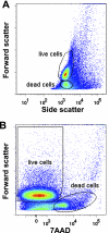
Fig. 1.
Fig. 1.
Discrimination of live and dead cells by forward scatter (FSC), side scatter (SSC), and 7-amino-actinomycin D (7AAD). A: analysis of splenocytes (erythrocyte free) using FSC and SSC. Upper population denoted as “live cells” represent viable splenocytes. Bottom population denoted as “dead cells” represent nonviable cells, which should be excluded from the sorting gate when preparing to sort for CD4+CD45RBhigh/low cells. B: viability of the cells can also be assessed by staining with 7AAD. In this graph only nonviable cells stain with 7AAD. Data were acquired by using a BD LSR II flow cytometer and analyzed with FlowJo software (ver. 7.2.2 for PC).
Fig. 2.
Fig. 2.
Body weight following adoptive transfer of CD4+CD45RBhigh or CD4+CD45RBlow T cells from wild-type donors into RAG−/− recipients. CD4+CD45RBhigh (•) or CD4+CD45RBlow T cells (○; 0.5 × 10⁶ cells of each) were injected [intraperitoneally (ip)] into RAG−/− mice to induce chronic colitis. Note the gradual loss of body weight beginning at week 4 for the mice reconstituted with CD4+CD45RBhigh T cells. Data derived from Ref. .
Fig. 3.
Fig. 3.
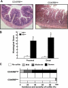
Histopathology of chronic colitis. A: representative micrographs of RAG−/− mice reconstituted with either CD4+CD45RBhigh (RBhigh) or CD4+CD45RBlow (RBlow) T cells (0.5 × 10⁶ cells) at 8 wk posttransfer. Note the large increase in bowel wall thickness, inflammatory cell infiltrate, and the appearance of crypt abscess (arrow) in the RAG−/− reconstituted with CD4+CD45RBhigh T cells. B: blinded histopathology scores of colons from both groups of mice (n = 9 for each group). See methods for scoring criteria. C: incidence and severity of chronic colitis. Animals receiving a histopathology score of 0 were judged to be without disease (no colitis); mild colitis received histopathology scores of 1–5; moderate colitis received histopathology scores of 6–10, and severe colitis received histopathology scores 11–17. We have found that >80% of the RAG−/− injected with CD4+CD45RBhigh T cells develop moderate-to-severe colitis at 8 wk following adoptive transfer.
Fig. 4.
Fig. 4.
High magnification of colon obtained from RAG−/− at 8 wk following adoptive transfer with CD4+CD45RBhigh T cells. Note the large mixed leukocyte infiltrate composed primarily of granulocytes, T cells, and monocytes (inset).
Fig. 5.
Fig. 5.
Correlation between colonic weight-to-length (g/cm) ratio and blinded histopathological scores. ▴, RAG−/− mice reconstituted with CD4+CD45RBhigh T cells (n = 36); ▿, RAG−/− mice reconstituted with CD4+CD45RBlow T cells (n = 7); •, RAG−/− mice reconstituted with both CD4+CD45RBhigh and CD4+CD45RBlow T cells (n = 14). Data derived from Ref. .
Fig. 6.
Fig. 6.
Intracellular staining for interleukin-17 (IL-17) and interferon-γ (IFN-γ) in T cells obtained from the mesenteric lymph nodes (MLNs) and colonic lamina propria (colon LP) of RAG−/− mice reconstituted with CD4+CD45RBhigh T cells at 8 wk posttransfer. Mononuclear cells were prepared from MLN and colon LP as previously described (17, 18). Cells were cultured for 16 h in a CD3-coated 24-well plate with soluble CD28 (1 μg/ml final) in complete RPMI-1640 medium. GolgiStop was added for the last 6 h. Cells were harvested, stained with anti-CD4, permeabilized by use of the BD cytofix/cytoperm kit and stained with isotype control monoclonal antibody (mAb) or IL-17 and IFN-γ mAbs. Contour plots are shown gated on CD4+ cells. Data were acquired by BD LSR II flow cytometer and analyzed using FlowJo software (ver. 7.2.2 for PC).
Fig. 7.
Fig. 7.
Histopathology, incidence, and severity of chronic colitis of RAG−/− mice reconstituted with CD4+CD45RBhigh T cells and treated with either an irrelevant isotype monoclonal antibody (cmAb) or a mAb against mouse tumor necrosis factor-α (TNF mAb). At 48 h following reconstitution of RAG−/− with T cells, mice were treated with 0.5 mg of either mAb (ip) 3 times per week for 8 wk.
Fig. 8.
Fig. 8.
Histopathology, incidence, and severity of jejunal (A) and ileal (B) inflammation in RAG−/− mice reconstituted with CD4+CD45RBhigh or CD4+CD45RBlow T cells. Note the villus blunting, bowel wall thickening, and inflammatory infiltrate in the mice injected with CD4+CD45RBhigh T cells. Incidence and severity of inflammation were based on scores described in the Fig. 3 legend. Data derived from Ref. .
Fig. 9.
Fig. 9.
Liver histopathology in RAG−/− mice reconstituted with CD4+CD45RBhigh T cells at 8 wk posttransfer. Note the periportal and intralobular inflammatory infiltrate composed primarily of mononuclear cells.
Fig. 10.
Fig. 10.
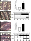
Adoptive transfer of CD11a-deficient (CD11a−/−) CD4+CD45RBhigh T cells into RAG-1−/− recipients fails to induce colitis. Histopathology, incidence, and severity of colitis were determined at 8 wk posttransfer. Data obtained from Ref. .
Fig. 11.
Fig. 11.
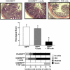
Cotransfer of CD11a−/− regulatory T cells (Tregs; CD4+CD25+) with CD4+CD45RBhigh T cells suppresses the development of chronic colitis as effectively as wild-type Tregs. Wild-type or CD11a−/− Tregs (0.25 × 10⁶ cells) were injected into RAG−/− recipients at 2 wk following injection of CD4+CD45RBhigh T cells (0.5 × 10⁶ cells). Histopathology was quantified at 8 wk posttransfer.
Fig. 12.
Fig. 12.
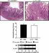
Adoptive transfer of CD4+ T cells obtained from interleukin-10-deficient (IL-10−/−) mice induces chronic colitis in RAG−/− recipients at 8 wk posttransfer. IL-10−/− splenocytes were enriched for CD4+ T cells (>85%) using a commercially available negative selection kit specifically for CD4+ T cells (Dynal). Wild-type (WT) CD4+CD45RBhigh T cells were purified by flow cytometry as described in the methods. RAG-1−/− recipients were injected (ip) either with IL-10−/− CD4+ T cells (1 × 10⁶ cells) or with CD4+CD45RBhigh T cells (0.5 × 10⁶ cells). Note that IL-10−/− CD4+ injected mice possessed a similar incidence and severity of chronic colitis as did RAG−/− mice injected with WT naive T cells purified by flow cytometry. n = 8 for each group.

References

    1. Berg DJ, Davidson N, Kuhn R, Muller W, Menon S, Holland G, Thompson-Snipes L, Leach MW, Rennick D. Enterocolitis and colon cancer in interleukin-10-deficient mice are associated with aberrant cytokine production and CD4 (+) TH1-like responses. J Clin Invest 98: 1010–1020, 1996. - PMC - PubMed
    1. Coombes JL, Robinson NJ, Maloy KJ, Uhlig HH, Powrie F. Regulatory T cells and intestinal homeostasis. Immunol Rev 204: 184–194, 2005. - PubMed
    1. Dohi T, Fujihashi K, Koga T, Shirai Y, Kawamura YI, Ejima C, Kato R, Saitoh K, McGhee JR. T helper type-2 cells induce ileal villus atrophy, goblet cell metaplasia, and wasting disease in T cell-deficient mice. Gastroenterology 124: 672–682, 2003. - PubMed
    1. Elson CO, Cong Y, McCracken VJ, Dimmitt RA, Lorenz RG, Weaver CT. Experimental models of inflammatory bowel disease reveal innate, adaptive, and regulatory mechanisms of host dialogue with the microbiota. Immunol Rev 206: 260–276, 2005. - PubMed
    1. Fiocchi C Inflammatory bowel disease: etiology and pathogenesis. Gastroenterology 115: 182–205, 1998. - PubMed

Publication types

MeSH terms

LinkOut - more resources