. 2009 Jan;119(1):193-202.
doi: 10.1172/JCI36983. Epub 2008 Dec 1.

IRBIT coordinates epithelial fluid and HCO3- secretion by stimulating the transporters pNBC1 and CFTR in the murine pancreatic duct

Affiliations

IRBIT coordinates epithelial fluid and HCO3- secretion by stimulating the transporters pNBC1 and CFTR in the murine pancreatic duct

Dongki Yang et al. J Clin Invest. 2009 Jan.

Abstract

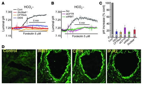
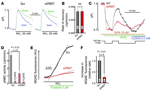
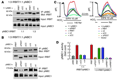
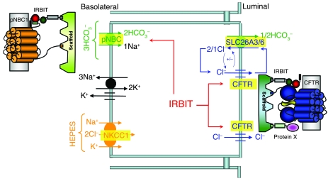
Fluid and HCO3- secretion are vital functions of secretory epithelia. In most epithelia, this entails HCO3- entry at the basolateral membrane, mediated by the Na+-HCO3- cotransporter, pNBC1, and exit at the luminal membrane, mediated by a CFTR-SLC26 transporters complex. Here we report that the protein IRBIT (inositol-1,4,5-trisphosphate [IP3] receptors binding protein released with IP3), a previously identified activator of pNBC1, activates both the basolateral pNBC1 and the luminal CFTR to coordinate fluid and HCO3- secretion by the pancreatic duct. We used video microscopy and ion selective microelectrodes to measure fluid secretion and Cl- and HCO3- concentrations in cultured murine sealed intralobular pancreatic ducts. Short interference RNA-mediated knockdown of IRBIT markedly inhibited ductal pNBC1 and CFTR activities, luminal Cl- absorption and HCO3- secretion, and the associated fluid secretion. Single-channel measurements suggested that IRBIT regulated CFTR by reducing channel mean close time. Furthermore, expression of IRBIT constructs in HEK cells revealed that activation of pNBC1 required only the IRBIT PEST domain, while activation of CFTR required multiple IRBIT domains, suggesting that IRBIT activates these transporters by different mechanisms. These findings define IRBIT as a key coordinator of epithelial fluid and HCO3- secretion and may have implications to all CFTR-expressing epithelia and to cystic fibrosis.


Figures

Figure 1
Figure 1. Knockdown of IRBIT inhibits pancreatic duct fluid secretion.
(A) RT-PCR of IRBIT mRNA in sealed ducts treated with scrambled (Scr) or 3 IRBIT dicer siRNA is shown. (B) Fluid secretion in ducts perfused in HCO3-buffered media and treated with scrambled (filled circles) or IRBIT siRNA (open circles) and stimulated with 5 μM forskolin is shown. Fluid secretion in C and D was measured as in B, except that the ducts were perfused with HEPES-buffered media. (C) The ducts were treated with IRBIT dicer siRNA (open circles). (D) The ducts were treated with CFTR dicer siRNA (open triangles) or incubated with 10 μM of CFTRinh-172 (CFTRinh, open circles). The results are given as the mean ± SEM of 5–8 experiments.
Figure 2
Figure 2. Properties of Cl transport by the pancreatic duct.
(A) A bright-field image of a pancreatic duct held in place by a holding pipette and impaled with a Cl recording electrode is shown. Original magnification, ×100. (B) The changes in luminal Cl of sealed ducts perfused with HEPES-buffered (green trace) or HCO3-buffered media (blue trace) are shown. (C) The changes in luminal Cl concentration after 45 seconds and 5 minutes of stimulation with 5 μM forskolin are summarized (mean ± SEM). (D) The changes in luminal Cl of sealed ducts perfused with HCO3-buffered media (black trace), containing 0.5 mM DIDS to inhibit pNBC1 (blue trace), or treated with CFTR (green trace) or IRBIT (red trace) siRNA are shown. (E) Summary (mean ± SEM) of Cl secretory activity, as in D. Numbers within bars denote number of experiments. (F) The changes in luminal Cl of sealed ducts perfused with HEPES-buffered media (black trace), containing 0.5 mM DIDS (blue trace), 10 μM CFTRinh-172 (green trace), or 0.1 mM Bumetanide (Bume; brown trace) to inhibit NKKC1 are shown. (G) The changes in luminal Cl of sealed ducts perfused with HEPES-buffered media and treated with scrambled (black trace), CFTR (green trace), or IRBIT (red trace) siRNA are shown. (H) Summary (mean ± SEM) of Cl secretory activity, as in F. Numbers within bars denote number of experiments. Cont, control.
Figure 3
Figure 3. Properties of HCO3 secretion by the pancreatic duct.
(A) The changes in luminal pH of sealed ducts perfused with HCO3-buffered media (black trace), containing 0.5 mM DIDS (blue trace), or 10 μM CFTRinh-172 (orange trace) are shown. The red trace shows luminal pH of sealed ducts from Slc26a6–/– mice. (B) The changes in luminal pH of sealed ducts perfused with HCO3-buffered media and treated with scrambled (black trace), CFTR (purple trace), or IRBIT (green trace) siRNA are shown. (C) Summary (mean ± SEM) of increase in pHi, as in A. Numbers within bars denote number of experiments. (D) Immunolocalization of IRBIT, CFTR and IP3R2. The first image is the control in which primary antibodies were omitted. Original magnification, ×1,000.
Figure 4
Figure 4. IRBIT stimulates pancreatic duct pNBC1 and CFTR activity.
(A) Measurement of pHi in ducts treated with scrambled (left traces) or IRBIT siRNA (right traces) and perfused with HEPES-buffered media is shown. Where indicated the ducts were exposed to media in which 20 mM NH4Cl replaced 20 mM NaCl, with and without 100 μM bumetanide, as indicated. The inhibition of pHi recovery by bumetanide is given in B in the form of ΔpH/min (mean ± SEM). (C) All solutions contained 10 μM S-(N-ethyl-N-isopropyl) amiloride (EIPA). Ducts treated with scrambled (black trace) or IRBIT siRNA (red trace) in HEPES-buffered media were perfused with Na+-free HCO3-buffered media. After completion of the acidification, the ducts were perfused with media containing 140 mM Na+ and then were perfused again with Na+-free media to reacidify the ducts. The ducts were used to measure recovery from acidification in the presence of the pNBC1 inhibitor 0.2 mM H2DIDS. The rates of Na+-dependent and H2DIDS-inhibitable changes in pHi reflect pNBC1 activity and are summarized in D. The results are given as mean ± SEM of 4 experiments. (E) The changes in MQAE fluorescence of ducts treated with scrambled (black trace) or IRBIT siRNA (red trace) and perfused in HEPES-buffered media are shown. Where indicated, the ducts were stimulated with forskolin and then perfused with media, in which all Cl was replaced with NO3. The dashed lines mark the largest slopes. (F) Summary (mean ± SEM) of increase in MQAE fluorescence, as in E. Numbers within bars denote number of experiments.
Figure 5
Figure 5. IRBIT activates CFTR.
(A) The Cl current in HEK cells transfected with CFTR (open circles) or CFTR and IRBIT (filled circles) in a 1:3 ratio is shown. Where indicated, the cells were stimulated with forskolin and treated with CFTRinh-172. (B) Summary of current density in cells expressing CFTR (green) or CFTR and IRBIT in 1:1 (blue) or 1:3 (red) ratios are shown. Results are given as the mean ± SEM of the number of experiments indicated in the columns. (C) The surface expression of CFTR in cells transfected with vector (control), CFTR, or CFTR and IRBIT in a 1:3 ratio, as indicated, is shown. (D) Example traces of single CFTR channel activity are shown (c, close channel state; o, open channel state), and (EG) show the (mean ± SEM) of (E) the open probability (NPo), (F) burst duration, and (G) interburst duration.
Figure 6
Figure 6. Role of IRBIT domains in interaction with and activation of CFTR.
(A) A model of the IRBIT domains and the coimmunoprecipitation of IRBIT and CFTR is shown. HEK cells were transfected with a 1:1 ratio of CFTR and the IRBIT truncation mutants or the IRBIT mutants alone (controls) and were used to IP CFTR and blot for IRBIT (upper blots) or IP IRBIT and blot for CFTR (lower blots). ΔCC, IRBITΔCC; ΔPEST, IRBITΔPEST; ΔCCΔ4, IRBITΔCCΔ4. (B) Example traces of CFTR current in HEK cells transfected with CFTR and the indicated IRBIT mutants at a 1:3 ratio are shown. The turquoise trace in the right is from a cell treated with IRBIT siRNA. (C) The current density calculated in pA/pF (mean ± SEM). The number of experiments is listed on the columns. (D) The coimmunoprecipitation of IRBIT and CFTR in cells transfected either with CFTR or with CFTRΔ4 and the indicated IRBIT constructs at a 1:3 ratio are shown. (E) Example traces of current measured in cells transfected with CFTRΔ4 or CFTRΔ4 and IRBIT at a 1:3 ratio. (F) The current density calculated in pA/pF (mean ± SEM). The number of experiments is listed on the columns.
Figure 7
Figure 7. Role of IRBIT domains in interaction with and activation of pNBC1.
(A and B) The coimmunoprecipitation of pNBC1 and the IRBIT truncation expressed at a 1:1 (A) or 1:3 (B) ratio. –, expressing only pNBC1. (C) The effect of IRBIT and the truncation mutants on pNBC1 activity when expressed in a 1:1 (left traces) or 1:3 (right traces) ratio. The turquoise trace is cells treated with IRBIT siRNA and transfected with pNBC1. (D) The fold increase of decrease in pNBC1 activity (mean ± SEM).
Figure 8
Figure 8. IRBIT coordinates pancreatic duct fluid and HCO3 secretion.
The model shows the key transporters involved in pancreatic duct fluid and HCO3 secretion. IRBIT forms complexes with the BLM pNBC1 and the LM CFTR that are assembled by PDZ domains-containing scaffolding proteins. Activation of pNBC1 and CFTR by IRBIT serves to coordinate HCO3 entry at the BLM and HCO3 exit at the LM and ensures the fidelity of epithelial fluid and HCO3 secretion.

References

    1. Durie P.R. The pathophysiology of the pancreatic defect in cystic fibrosis. Acta Paediatr. Scand. Suppl. 1989;363:41–44. - PubMed
    1. Baron J.H. The pancreas. Mt. Sinai J. Med. 2000;67:68–75. - PubMed
    1. Steward M.C., Ishiguro H., Case R.M. Mechanisms of bicarbonate secretion in the pancreatic duct. Annu. Rev. Physiol. 2005;67:377–409. doi: 10.1146/annurev.physiol.67.031103.153247. - DOI - PubMed
    1. Ishiguro H., Steward M.C., Lindsay A.R., Case R.M. Accumulation of intracellular HCO3- by Na(+)-HCO3- cotransport in interlobular ducts from guinea-pig pancreas. J. Physiol. 1996;495:169–178. - PMC - PubMed
    1. Zhao H., Star R.A., Muallem S. Membrane localization of H+ and HCO3- transporters in the rat pancreatic duct. J. Gen. Physiol. 1994;104:57–85. doi: 10.1085/jgp.104.1.57. - DOI - PMC - PubMed

Publication types

MeSH terms