. 2008 Dec;118(12):3943-53.
doi: 10.1172/JCI37156. Epub 2008 Nov 6.

Proliferation of human HCC cells and chemically induced mouse liver cancers requires JNK1-dependent p21 downregulation

Affiliations

Proliferation of human HCC cells and chemically induced mouse liver cancers requires JNK1-dependent p21 downregulation

Lijian Hui et al. J Clin Invest. 2008 Dec.

Abstract

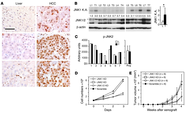
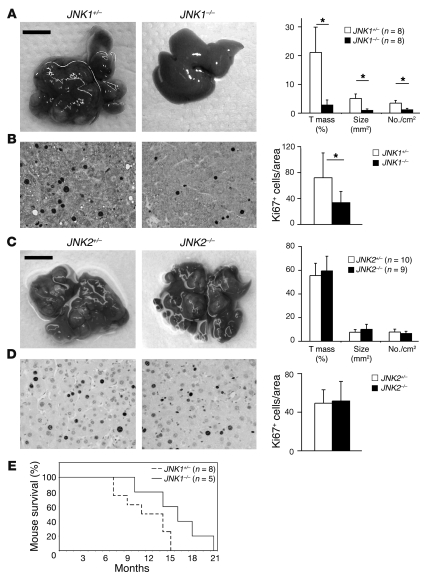
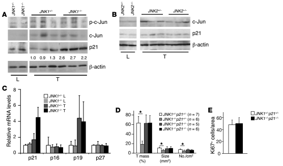
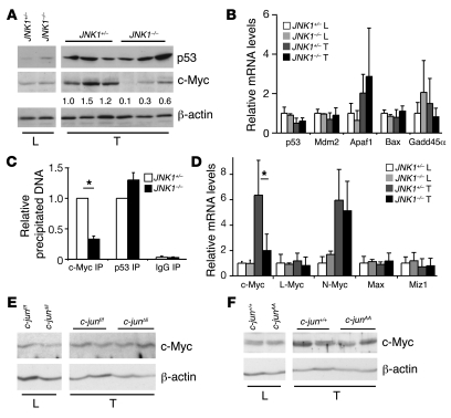
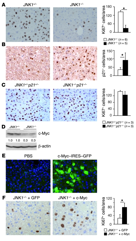
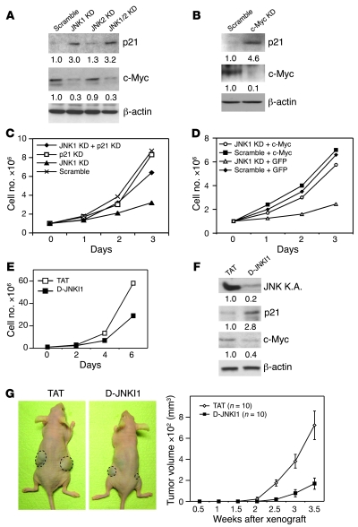
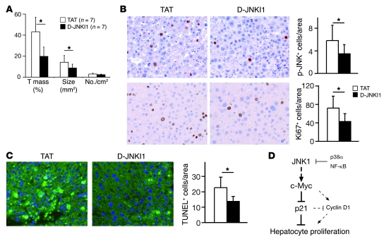
JNK proteins have been shown to be involved in liver carcinogenesis in mice, but the extent of their involvement in the development of human liver cancers is unknown. Here, we show that activation of JNK1 but not JNK2 was increased in human primary hepatocellular carcinomas (HCCs). Further, JNK1 was required for human HCC cell proliferation in vitro and tumorigenesis after xenotransplantation. Importantly, mice lacking JNK1 displayed decreased tumor cell proliferation in a mouse model of liver carcinogenesis and decreased hepatocyte proliferation in a mouse model of liver regeneration. In both cases, impaired proliferation was caused by increased expression of p21, a cell-cycle inhibitor, and reduced expression of c-Myc, a negative regulator of p21. Genetic inactivation of p21 in JNK1-/- mice restored hepatocyte proliferation in models of both liver carcinogenesis and liver regeneration, and overexpression of c-Myc increased proliferation of JNK1-/- liver cells. Similarly, JNK1 was found to control the proliferation of human HCC cells by affecting p21 and c-Myc expression. Pharmacologic inhibition of JNK reduced the growth of both xenografted human HCC cells and chemically induced mouse liver cancers. These findings provide a mechanistic link between JNK activity and liver cell proliferation via p21 and c-Myc and suggest JNK targeting can be considered as a new therapeutic approach for HCC treatment.


Figures

Figure 1
Figure 1. JNK1 activity is increased in human HCC.
(A) Immunohistochemical staining of p-JNK on a tissue microarray of paired human HCCs and nonneoplastic liver tissues. p-JNK staining in HCCs was significantly stronger than that in nonneoplastic liver tissues in 23 out of 41 matched pairs. P < 0.05, Wilcoxon-Mann-Whitney test. Three representative paired samples are shown. Cells with activated JNK show brown nuclear staining. Scale bar: 50 μm. (B) Kinase activities of JNK1 were determined in paired samples of human nonneoplastic livers and HCCs by immunocomplex kinase assay using GST–c-Jun as substrate. Total JNK and β-actin levels were assayed by Western blot. L, liver; T, tumor. Numbers between blots denote JNK kinase activity (K.A.), quantified by normalizing to β-actin levels. Average of JNK1 activities of all samples is shown in the right panel. *P < 0.05, Student’s paired t test. (C) p-JNK2 levels in human HCC samples were analyzed by ELISA assay using total cell extraction. Average of p-JNK2 levels is shown in the right panel. (D) Huh7 cells were infected with lentiviruses expressing shRNAs against JNK1, JNK2, and JNK1/2 as well as scramble control shRNA. Proliferation of cultured cells was analyzed using cells at passage 2 after virus infection. One representative experiment of 3 is shown. KD, knockdown. (E) Tumor formation of JNK1, JNK2, JNK1/2 knockdown, and scramble control Huh7 cells were analyzed after these cells were subcutaneously implanted in nude mice. Data are expressed as mean ± SD.
Figure 2
Figure 2. JNK1 but not JNK2 enhances proliferation of mouse liver cancer cells.
(A) DEN-induced liver cancers in JNK1+/– and JNK1–/– mice are shown. Quantification of tumor mass (percentage of total tumor size compared with total liver tissue size), average tumor size (mm2), and tumor numbers per cm2 on H&E-stained liver sections are shown in the right panel. (B) Proliferation of liver cancer cells in JNK1+/– and JNK1–/– mice was analyzed by immunostaining for Ki67. Ki67-positive cells are indicated by a brown-stained nucleus. Ki67-positive cells in cancers were quantified. In total, 21 liver tumors from 6 JNK1+/– mice and 15 tumors from 5 JNK1–/– mice were analyzed. *P < 0.05, Student’s t test. (C) Liver cancers from DEN-treated JNK2+/– and JNK2–/– mice are shown. Quantification of tumor mass, average tumor size, and tumor numbers are shown in the right panel. Scale bars: 1 cm (A and C). (D) Proliferation of liver cancer cells in JNK2+/– and JNK2–/– mice was determined by Ki67 staining. Ki67-positive cells were quantified. Original magnification, ×200 (B and D). (E) Kaplan-Meier survival curve of JNK1+/– and JNK1–/– mice subjected to DEN-Pb liver carcinogenesis protocol. P < 0.05, Gehan’s test. Data are expressed as mean ± SD.
Figure 3
Figure 3. Upregulation of p21 causes reduced proliferation of JNK1–/– liver cancer cells.
(A) Protein levels of p–c-Jun, c-Jun, and p21 were determined by Western blot in JNK1+/– and JNK1–/– liver and cancer tissues. β-actin levels were used as loading control. Numbers below p21 blot represent quantification as normalized to β-actin. (B) c-Jun and p21 protein levels were determined by Western blot in JNK2+/– and JNK2–/– liver cancer tissues. β-actin was used as loading control. (C) mRNA levels of p21, p16, p19, and p27 were determined by qRT-PCR in cancer and normal liver tissues from JNK1+/– and JNK1–/– mice. (D) DEN-induced liver cancers in JNK1+/–p21+/–, JNK1–/–p21+/–, JNK1+/–p21–/–, and JNK1–/–p21–/– mice were quantified in terms of tumor mass, average tumor size, and tumor numbers. (E) Proliferation of cancer cells was analyzed by Ki67 staining on liver sections from DEN-treated JNK1+/–p21+/– and JNK1–/–p21–/– mice. Quantification of Ki67-positive cells in cancers is shown. *P < 0.05, Student’s t test. Data are expressed as mean ± SD.
Figure 4
Figure 4. c-Myc expression levels are reduced in JNK1–/– liver cancers.
(A) Protein levels of p53 and c-Myc were analyzed by Western blots in JNK1–/– cancer and normal liver tissues. β-actin levels were used as loading control. c-Myc levels were quantified. (B) mRNA levels of p53 and its target genes Mdm2, Apaf1, Bax, and Gadd45α were determined by qRT-PCR in JNK1+/– and JNK1–/– liver and cancer tissues. (C) The binding of c-Myc and p53 to the p21 promoter (–216 to –108) was analyzed in pooled DEN-induced liver cancers using ChIP assays (c-Myc IP and p53 IP). Rabbit IgG was used as negative control (IgG IP). The amount of c-Myc or p53 binding p21 promoter DNA was quantified by qRT-PCR assays. (D) mRNA levels of c-Myc, L-Myc, N-Myc, Max, and Miz1 were analyzed by qRT-PCR in JNK1–/– liver cancers. (E and F) c-Myc protein levels were analyzed by Western blot in liver cancers from c-junΔli and c-junAA mice. *P < 0.05, Student’s t test. Data are expressed as mean ± SD.
Figure 5
Figure 5. JNK1 facilitates hepatocyte proliferation by regulating expression of p21 and c-Myc.
(A) Proliferating hepatocytes were analyzed by Ki67 staining in JNK1–/– livers 48 hours after partial hepatectomy. Quantification of Ki67-positive cells is shown. (B) Immunohistochemical staining of p21 on liver sections of JNK1+/– and JNK1–/– mice during liver regeneration (brown nuclear staining). (C) Hepatocyte proliferation in JNK1+/–p21+/– and JNK1–/–p21–/– mice was analyzed by Ki67 immunostaining at 48 hours after partial hepatectomy. (D) c-Myc protein levels were analyzed by Western blot in livers from JNK1+/– and JNK1–/– mice after partial hepatectomy. (E) Mice were injected with PBS or 30 μg of c-Myc–IRES–GFP bicistronic plasmid through tail veins. Two days after injection, transfection of hepatocytes was monitored by GFP expression in liver cryosections. Nuclei were stained blue by DAPI. (F) Hepatocyte proliferation was analyzed 48 hours after partial hepatectomy in JNK1–/– mice with hydrodynamics-based GFP or c-Myc–IRES–GFP (c-Myc) transfection. *P < 0.05, Student’s t test. Original magnification, ×200. Data are expressed as mean ± SD.
Figure 6
Figure 6. JNK1 regulates p21 and c-Myc expression in human Huh7 HCC cells.
(A) p21 and c-Myc protein levels in JNK1, JNK2, and JNK1/2 knockdown Huh7 cells and scramble controls were determined by Western blot. (B) p21 and c-Myc levels were assayed by Western blot in human Huh7 cells transfected with scramble control siRNA or c-Myc siRNA. Quantification is normalized to β-actin levels. (C) Proliferation rate of Huh7 cells with both JNK1 and p21 knockdown was analyzed. (D) Huh7 cells with scramble or JNK1 shRNAs were transfected with either pIRES-GFP (GFP) or c-Myc–IRES2–AcGFP (c-Myc). Proliferation of these cells was analyzed for 3 days. (E) Proliferation of cultured Huh7 cells was reduced upon treatment with D-JNKI1 (20 μM). One representative experiment of 3 is shown. (F) JNK kinase activity in D-JNKI1–treated (20 μM) Huh7 cultures was analyzed by immunocomplex kinase assay using GST–c-Jun as substrates. p21 and c-Myc protein levels were determined by Western blot. Quantification was normalized to β-actin. (G) Tumor growth of subcutaneously implanted Huh7 cells was analyzed following 4 weeks of treatment with TAT or D-JNKI1 (5 mg/kg) in nude mice. Data are expressed as mean ± SD.
Figure 7
Figure 7. JNK inhibitor treatment reduces liver cancer development.
(A) Five months after DEN injection, mice were treated with JNK inhibitor peptide D-JNKI1 or control peptide TAT (5 mg/kg) twice a week for 3 months. Quantification of tumor mass, tumor size, and tumor number from D-JNKI1– or TAT-treated mice is shown. (B) Phosphorylation of JNK in DEN-induced liver cancers from D-JNKI1– or TAT-treated mice was analyzed by immunohistochemical staining on liver sections (brown nuclear staining). Proliferation of cancer cells was analyzed by immunostaining for Ki67. (C) Wild-type mice at 4 weeks of age were injected with D-JNKI1 or TAT at 5 mg/kg 2 days before DEN treatment and killed 24 hours after. DEN-induced cell death was analyzed by TUNEL staining on liver sections. TUNEL-positive cells (green-stained nucleus) were quantified. *P < 0.05, Student’s t test. Original magnification, ×200. (D) Schematic model of JNK1-dependent hepatocyte proliferation. JNK1 activation and decreased p21 levels are apparently important for cell proliferation during liver carcinogenesis and regeneration. p21, repressed by c-Myc, attenuates proliferation of liver cells, probably via suppressing cyclin D1. Importantly, this pathway is negatively regulated by p38α and NF-κB stress-signaling pathways and appears to be independent of c-Jun. Data are expressed as mean ± SD.

References

    1. Farazi P.A., DePinho R.A. Hepatocellular carcinoma pathogenesis: from genes to environment. Nat. Rev. Cancer. 2006;6:674–687. doi: 10.1038/nrc1934. - DOI - PubMed
    1. Eferl R., et al. Liver tumor development. c-Jun antagonizes the proapoptotic activity of p53. Cell. 2003;112:181–192. doi: 10.1016/S0092-8674(03)00042-4. - DOI - PubMed
    1. Hui L., et al. p38alpha suppresses normal and cancer cell proliferation by antagonizing the JNK-c-Jun pathway. Nat. Genet. 2007;39:741–749. doi: 10.1038/ng2033. - DOI - PubMed
    1. Maeda S., Kamata H., Luo J.L., Leffert H., Karin M. IKKbeta couples hepatocyte death to cytokine-driven compensatory proliferation that promotes chemical hepatocarcinogenesis. Cell. 2005;121:977–990. doi: 10.1016/j.cell.2005.04.014. - DOI - PubMed
    1. Sakurai T., Maeda S., Chang L., Karin M. Loss of hepatic NF-kappa B activity enhances chemical hepatocarcinogenesis through sustained c-Jun N-terminal kinase 1 activation. Proc. Natl. Acad. Sci. U. S. A. 2006;103:10544–10551. doi: 10.1073/pnas.0603499103. - DOI - PMC - PubMed

Publication types

MeSH terms

Substances