. 2009 Jan;87(1):25-30.
doi: 10.1007/s00109-008-0422-3. Epub 2008 Nov 26.

RNaseH2 mutants that cause Aicardi-Goutieres syndrome are active nucleases

Affiliations

RNaseH2 mutants that cause Aicardi-Goutieres syndrome are active nucleases

Fred W Perrino et al. J Mol Med (Berl). 2009 Jan.

Abstract

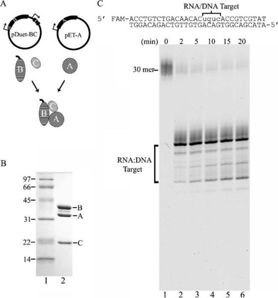
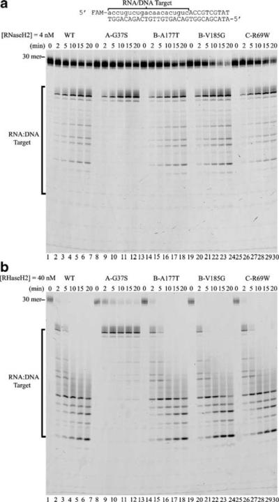
Mutations in the genes encoding the RNaseH2 and TREX1 nucleases have been identified in patients with Aicardi-Goutieres syndrome (AGS). To determine if the AGS RNaseH2 mutations result in the loss of nuclease activity, the human wild-type RNaseH2 and four mutant complexes that constitute the majority of mutations identified in AGS patients have been prepared and tested for ribonuclease H activity. The heterotrimeric structures of the mutant RNaseH2 complexes are intact. Furthermore, the ribonuclease H activities of the mutant complexes are indistinguishable from the wild-type enzyme with the exception of the RNaseH2 subunit A (Gly37Ser) mutant, which exhibits some evidence of altered nuclease specificity. These data indicate that the mechanism of RNaseH2 dysfunction in AGS cannot be simply explained by loss of ribonuclease H activity and points to a more complex mechanism perhaps mediated through altered interactions with as yet identified nucleic acids or protein partners.


Figures

Fig. 1
Fig. 1
The human RNaseH2 is an active heterotrimeric ribonuclease H. A His6-tag was engineered at the N terminus (hatched marks)of the RNASEH2B gene and cloned upstream of the untagged RNASEH2C gene to yield the pDuet-BC plasmid (a). The untagged RNASEH2A gene was cloned on a separate plasmid to yield pET-A (a). Expression of each gene is controlled by a T7 promoter (arrows). The purified recombinant RNaseH2 three-subunit complex (6 μg) was subjected to 15% sodium dodecyl sulfate polyacrylamide gel electrophoresis (SDS-PAGE), and the gel was stained with Coomassie Brilliant Blue (b, lane 2). The positions of migration of the molecular weight standards (b, lane 1) and RNaseH2 subunits are indicated. An RNaseH2 (40 nM) reaction was prepared with the DNA16-RNA4-DNA10/DNA30 duplex, and samples were removed at the indicated times after incubation at 25°C. Reaction products were subjected to electrophoresis on a 23% urea-polyacrylamide gel (c). The positions of migration of the 30-mer and products (RNA/DNA Target) are indicated. The product sizes were determined by comparison to a ladder of fragments generated by TREX1 exonuclease digestion of the same oligomer and run in adjacent lanes (not shown)
Fig. 2
Fig. 2
The RNaseH2 mutants are active heterotrimeric ribonuclease Hs. Site-directed mutations were introduced into the RNASEH2A, RNASEH2B, and RNASEH2C expression constructs, and the RNaseH2 mutant complexes were purified as described in “Materials and methods.” Approximately 6 μg of each RNaseH2 complex was subjected to 15% SDS-PAGE, and the gel was stained with Coomassie Brilliant Blue (a, lanes 26). The positions of migration of the molecular weight standards (a, lane 1) and RNaseH2 subunits are indicated. RNaseH2 (40 nM) reactions containing the indicated mutant enzyme were prepared with the DNA16-RNA4-DNA10/DNA30 duplex, and samples were removed at the indicated times after incubation at 25°C. Reaction products were subjected to electrophoresis on a 23% urea-polyacrylamide gel (b). The positions of migration of the 30-mer and products (RNA/DNA Target) are indicated. The product sizes were determined by comparison to a ladder of fragments generated by TREX1 exonuclease digestion of the same oligomer and run in adjacent lanes (not shown)
Fig. 3
Fig. 3
Altered specificity in the RNaseH2 subunit A Gly37Ser mutant. RNaseH2 reactions containing the indicated mutant enzyme (a, 4 nM) and (b, 40 nM) were prepared with the RNA20-DNA10/DNA30 duplex and samples were removed at the indicated times after incubation at 25°C. Reaction products were subjected to electrophoresis on 23% urea-polyacrylamide gels. The positions of migration of the 30-mer and products (RNA/DNA Target) are indicated. The product sizes were determined by comparison to a ladder of fragments generated by TREX1 exonuclease digestion of the same oligomer and run in adjacent lanes (not shown)

References

    1. Aicardi J, Goutieres F. A progressive familial encephalopathy in infancy with calcifications of the basal ganglia and chronic cerebrospinal-fluid lymphocytosis. Ann Neurol. 1984;15:49–54. - PubMed
    1. Goutieres F, Aicardi J, Barth PG, Lebon P. Aicardi–Goutieres syndrome: an update and results of interferon-alpha studies. Ann Neurol. 1998;44:900–907. - PubMed
    1. Goutieres F. Aicardi–Goutieres syndrome. Brain Dev. 2005;27:201–206. - PubMed
    1. Crow YJ, Hayward BE, Parmar R, et al. Mutations in the gene encoding the 3′–5′ DNA exonuclease TREX1 cause Aicardi–Goutieres syndrome at the AGS1 locus. Nat Genet. 2006;38:917–920. - PubMed
    1. Crow YJ, Leitch A, Hayward BE, et al. Mutations in genes encoding ribonuclease H2 subunits cause Aicardi–Goutieres syndrome and mimic congenital viral brain infection. Nat Genet. 2006;38:910–916. - PubMed

Publication types

MeSH terms