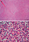
Non-traumatic splenic rupture: report of seven cases and review of the literature
- PMID: 19034976
- PMCID: PMC2773315
- DOI: 10.3748/wjg.14.6711
Non-traumatic splenic rupture: report of seven cases and review of the literature
Abstract
Aim: To evaluate seven patients with non-traumatic splenic rupture (NSR). NSR is an uncommon dramatic abdominal emergency that requires immediate diagnosis and prompt surgical treatment to ensure the patient's survival.
Methods: Within 11 years, seven cases were evaluated for patient characteristics, anamnesis and symptoms, method of diagnosis, findings of laparotomy, and etiology of NSR.
Results: There were six (86%) male and one female (14%) patient, whose mean age was 36 +/- 12.8 (17-56) years. We report here four cases of Plasmodium vivax malaria (cases I-IV), one case of hemodialysis (case V), one case of spontaneous splenic rupture (case VI), and one case of hairy cell leukemia (case VII). Splenectomy was performed in all patients. All of them made an uneventful recovery and were discharged in stable condition.
Conclusion: NSR is a rare entity that needs a high index of suspicion for diagnosis. Using ultrasonography or computer tomography, and peritoneal aspiration of fresh blood may assist in the diagnosis of NSR. Increased awareness of NSR can enhance early diagnosis and effective treatment.
Figures


References
-
- Hyun BH, Varga CF, Rubin RJ. Spontaneous and pathologic rupture of the spleen. Arch Surg. 1972;104:652–657. - PubMed
-
- Torricelli P, Coriani C, Marchetti M, Rossi A, Manenti A. Spontaneous rupture of the spleen: report of two cases. Abdom Imaging. 2001;26:290–293. - PubMed
-
- Andrews DF, Hernandez R, Grafton W, Williams DM. Pathologic rupture of the spleen in non-Hodgkin's lymphoma. Arch Intern Med. 1980;140:119–120. - PubMed
-
- Bauer TW, Haskins GE, Armitage JO. Splenic rupture in patients with hematologic malignancies. Cancer. 1981;48:2729–2733. - PubMed
-
- Gallerani M, Vanini A, Salmi R, Bertusi M. Spontaneous rupture of the spleen. Am J Emerg Med. 1996;14:333–334. - PubMed
Publication types
MeSH terms
LinkOut - more resources
Full Text Sources

