. 2009 Feb 15;124(4):853-61.
doi: 10.1002/ijc.24040.

CXCL8/IL-8 and CXCL12/SDF-1alpha co-operatively promote invasiveness and angiogenesis in pancreatic cancer

Affiliations

CXCL8/IL-8 and CXCL12/SDF-1alpha co-operatively promote invasiveness and angiogenesis in pancreatic cancer

Yoichi Matsuo et al. Int J Cancer. .

Abstract

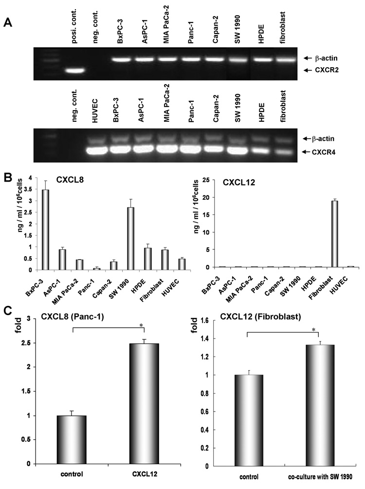
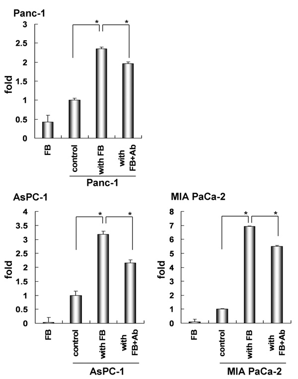
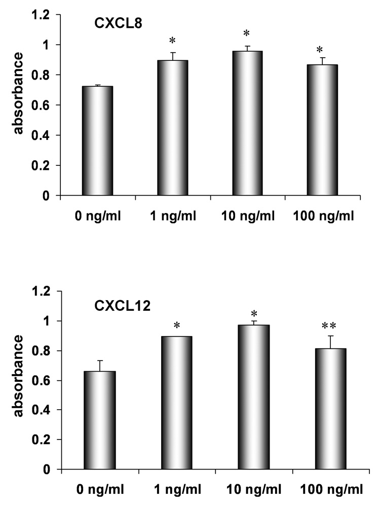
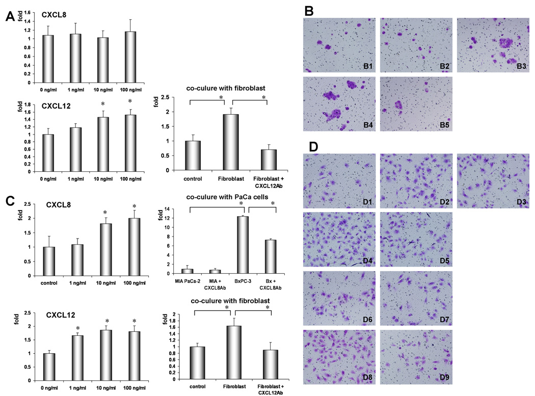
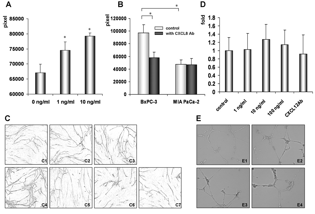
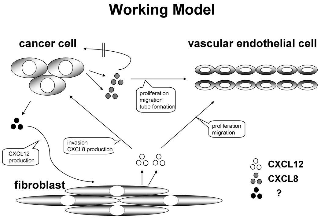
CXC-chemokines are involved in the chemotaxis of neutrophils, lymphocytes and monocytes. However, role of these chemokines in tumorigenesis, especially with regard to interaction between tumor and its microenvironment, has not been clearly elucidated. The purpose of this study was to analyze the co-operative role of CXCL8 and CXCL12 in the tumor-stromal interaction in pancreatic cancer (PaCa). Using enzyme-linked immunosorbent assay (ELISA) and reverse transcription polymerase chain reaction (RT-PCR), we initially confirmed the expression of ligands and receptors, respectively, of CXC-chemokines in PaCa and stromal cells. We examined the co-operative role of CXCL8 and CXCL12 in proliferation/invasion of PaCa and human umbilical vein endothelial cells (HUVECs), and in HUVEC tube-formations through tumor-stromal interaction by MTS, Matrigel invasion, and angiogenesis assays, respectively. We detected expression of CXCR4, but not CXCR2, in all PaCa cells and fibroblasts. PaCa cells secreted CXCL8, and fibroblast cells secreted CXCL12. CXCL8 production in PaCa was significantly enhanced by CXCL12, and CXCL12 production in fibroblasts was significantly enhanced by co-culturing with PaCa. CXCL8 enhanced proliferation/invasion of HUVECs but did not promote proliferation/invasion of PaCa. Both recombinant and PaCa-derived CXCL8 enhanced tube formation of HUVECs that were co-cultured with fibroblast cells. CXCL12 enhanced the proliferation/invasion of HUVECs and the invasion of PaCa cells but had no effect on tube formation of HUVEC. We showed that PaCa-derived CXCL8 and fibroblast-derived CXCL12 cooperatively induced angiogenesis in vitro by promoting HUVEC proliferation, invasion, and tube formation. Thus, corresponding receptors CXCR2 and CXCR4 are potential antiangiogenic and antimetastatic therapeutic targets in PaCa.


Figures

Figure 1
Figure 1. Expression of chemokine receptors and ligands in PaCa and stromal cells
(A) Detection of CXCR2 and CXCR4 mRNA in PaCa and stromal cells. PCR products were subjected to 1.2 % agarose gel electrophoresis and stained with ethidium bromide. (B) Quantification of CXCL8 and CXCL12 proteins in culture medium of PaCa and stromal cells. All cells were cultured for 48 h, and then the concentrations of both chemokines were measured by ELISA. (C) Change in chemokine production by tumor-stromal interaction. Alteration of CXCL8 production from PaCa (Panc-1) by stimulation with CXCL12 (20 ng/mL) (left) and alternation of CXCL12 production in fibroblasts by co-culturing with PaCa (SW 1990) (right). Values are expressed as mean ± SD. *, P < 0.01.
Figure 2
Figure 2. Effects on CXCL8 production from PaCa by co-culture with fibroblasts
The alteration of CXCL8 production from PaCa (Panc-1, AsPC-1, and MIA PaCa-2) by co-culturing with fibroblast (FB) was evaluated by ELISA as described in Materials and Methods. Also, to examine the role of fibroblast-derived CXCL12 on enhanced CXCL8 production from PaCa cell lines, PaCa cells were pre-treated with anti-CXCR4 Ab (Ab) for 1 h and ELISA experiments were performed as described in Materials and Methods. FB; fibroblast only, control; PaCa cells only, with FB; PaCa cells co-cultured with fibroblast, with FB + Ab; PaCa cells co-cultured with fibroblast treated with anti-CXCR4 Ab. Values are expressed as mean ± SD. *, P < 0.01.
Figure 3
Figure 3. Effects of CXCL8 and CXCL12 on proliferation of HUVECs
Cell proliferation was assessed using the MTS-assay as described in Material and Methods. Multiple comparisons were performed by one-way ANOVA followed by the SNK test. Values are expressed as mean ± SD. *, P < 0.01 and **, P < 0.05 versus control.
Figure 4
Figure 4. Effects of CXCL8 and CXCL12 on invasiveness of PaCa and HUVECs
Both PaCa invasiveness and HUVEC invasiveness were assessed by the BD Bio-Coat Matrigel invasion assay system (BD Biosciences) as described in Material and Methods. (A, B) PaCa invasion assay. PaCa (BxPC-3) invasion assay was performed under the condition of basal medium containing various concentrations of CXCL8 or CXCL12, or co-culture with fibroblast treated with or without anti-CXCL12 Ab (10µg/mL). Invading cells were fixed and stained with Diff-Quick stain. The invading cells were counted in five random microscopic fields (×200). Multiple comparisons were performed by one-way ANOVA followed by the SNK test. Bars indicate SD, *P < 0.01 compared with control. B1: control; B2: cultured with CXCL8 (10 ng/mL); B3: cultured with CXCL12 (10 ng/mL); B4: co-cultured with fibroblasts; B5: co-cultured with fibroblasts treated with anti-CXCL12 Ab (10 µg/mL). (C, D) HUVEC invasion assay. HUVEC invasion assay was performed under the condition of basal medium only or basal medium containing various concentrations of CXCL8 or CXCL12. To assess the interaction between HUVECs and PaCa or fibroblasts, HUVECs were co-cultured with PaCa (MIA PaCa-2 or BxPC-3) treated with or without anti-CXCL8 Ab or co-cultured with fibroblasts treated with or without anti-CXCL12 Ab. The invading cells were stained with Diff-Quick stain and counted in five random microscopic fields (×200). Multiple comparisons were performed by one-way ANOVA followed by the SNK test. Bars indicate SD, *P < 0.01 compared with control. D1: control; D2: cultured with CXCL8 (10 ng/mL); D3: cultured with CXCL12 (10 ng/mL); D4: co-cultured with BxPC-3; D5: co-cultured with BxPC-3 treated with anti-CXCL8 Ab (10 µg/mL); D6: co-cultured with MIA PaCa-2; D7: co-cultured with MIA PaCa-2 treated with anti-CXCL8 Ab (10 µg/mL); D8: co-cultured with fibroblast; D9: co-cultured with fibroblast treated with anti-CXCL12 Ab (10 µg/mL).
Figure 5
Figure 5. Effects of both chemokines on angiogenesis
(A) Effects of CXCL8 on tube formation by HUVEC. After incubation of the HUVEC/fibroblasts co-culture system (Kurabo Co.) in the presence or absence of CXCL8 for 11 days, HUVECs were stained with anti-CD31 antibody. Tube formation area was measured quantitatively using an image analyzer. Multiple comparisons were performed by one-way ANOVA followed by the SNK test. Bars indicate SD, *P < 0.01 compared with control. (B) Effects of PaCa cells with different concentrations of CXCL8 production on tube formation by HUVECs. BxPC-3 or MIA PaCa-2 cells were cultured with HUVECs and fibroblasts using a double chamber treated without anti-CXCL8 Ab (white columns) or with anti-CXCL8 Ab (black columns). The tube formation by HUVEC for each condition was measured using an image analyzer. Multiple comparisons were performed by one-way ANOVA followed by an SNK test. Bars indicate SD, *P < 0.01 compared with treatment with anti-CXCL8 Ab and MIA PaCa-2 cells. (C) Tube formation assay for angiogenesis by co-culturing PaCa, HUVEC, and fibroblasts. C1: control; C2: cultured with CXCL8 (1 ng/mL); C3: cultured with CXCL8 (10 ng/mL); C4: co-cultured with BxPC-3; C5: co-cultured with BxPC-3 treated with anti-CXCL8 Ab (10 µg/mL); C6: co-cultured with MIA PaCa-2; C7: co-cultured with MIA PaCa-2 treated with anti-CXCL8 Ab (10 µg/mL) (×40). (D) Effect of CXCL12 on HUVEC tube formation. Tube formation assay for angiogenesis on Matrigel was performed as described in Material and Methods. The number of endotubes was additionally quantified by counting 9 random fields/sample under the microscope (x100). (E) Tube formation assay for angiogenesis on Matrigel. E1: control, E2: cultured with CXCL12 (10 ng/mL); E3: cultured with CXCL12 (100 ng/mL); E4: cultured with anti-CXCL12 Ab (10 µg/mL).
Figure 6
Figure 6
CXCL8 and CXCL12 cooperatively promote invasion and angiogenesis of PaCa. PaCa cells secrete CXCL8. Fibroblast-derived CXCL12 enhanced CXCL8 production in PaCa cells. CXCL8 from PaCa up-regulated proliferation, invasion, and tube formation by HUVECs. Fibroblast cells produced CXCL12; this production was increased by co-culturing with PaCa cells. CXCL12 up-regulated not only PaCa invasion but also proliferation and invasion of HUVECs. In this way, CXCL8 and CXCL12 cooperatively promote invasion and angiogenesis of PaCa.

References

    1. Jemal A, Siegel R, Ward E, Murray T, Xu J, Thun MJ. Cancer statistics. CA Cancer J Clin. 2007;7:43–66. - PubMed
    1. Niedergethmann M, Alves F, Neff JK, Heidrich B, Aramin N, Li L, Pilarsky C, Grützmann R, Allgayer H, Post S, Gretz N. Gene expression profiling of liver metastases and tumour invasion in pancreatic cancer using an orthotopic SCID mouse model. Br J Cancer. 2007;16:1432–1440. - PMC - PubMed
    1. Murphy PM. Chemokines and the molecular basis of cancer metastasis. N Engl J Med. 2001;345:833–835. - PubMed
    1. Balkwill F, Mantovani A. Inflammation and cancer: back to Virchow? Lancet. 2001;357:539–545. - PubMed
    1. Homey B, Müller A, Zlotnik A. Chemokines: agents for the immunotherapy of cancer? Nat Rev Immunol. 2002;2:175–184. - PubMed

Publication types

MeSH terms

LinkOut - more resources