

Increased hydraulic conductance of human articular cartilage and subchondral bone plate with progression of osteoarthritis
- PMID: 19035476
- PMCID: PMC2603273
- DOI: 10.1002/art.24069
Increased hydraulic conductance of human articular cartilage and subchondral bone plate with progression of osteoarthritis
Abstract
Objective: Osteoarthritis (OA) is characterized by progressive degeneration of articular cartilage and remodeling of the subchondral bone plate, comprising calcified cartilage and underlying subchondral bone. Calcified cartilage remodeling due to upward invasion by vascular canals or to calcified cartilage erosion may contribute to biomechanical alteration of the osteochondral tissue and its subchondral bone plate component. The study hypothesis was that hydraulic conductance of osteochondral tissue and subchondral bone plate increases with structural changes indicative of increasing stages of OA.
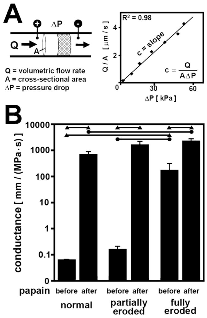
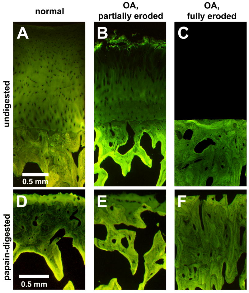
Methods: Osteochondral cores were harvested from the knees of cadaveric tissue donors and from discarded fragments from patients with OA undergoing knee surgery. The osteochondral cores from tissue donors were macroscopically normal, and the cores from patients with OA had partial-thickness or full-thickness erosion to bone. The cores were perfusion-tested to determine the hydraulic conductance, or ease of fluid flow, in their native state and after enzymatic removal of cartilage. Adjacent portions were analyzed by 3-dimensional histology for calcified cartilage, subchondral bone, and subchondral bone plate thickness and vascular canal density.
Results: Hydraulic conductance of native osteochondral tissue and subchondral bone plate was higher (2,700-fold and 3-fold, respectively) in fully eroded samples than in normal samples. The calcified cartilage layer was thicker (1.5-fold) in partially eroded samples than in normal samples but thinner and incomplete in fully eroded samples. Subchondral bone plate vascularity was altered with increasing stages of OA.
Conclusion: During joint loading, increased hydraulic conductance of the osteochondral tissue and subchondral bone plate could have deleterious biomechanical consequences for cartilage. Increased fluid exudation from overlying and opposing cartilage, increased fluid depressurization, and increased cartilage tissue strains could lead to chondrocyte death and cartilage damage.
Figures


References
-
- McCutchen CW. The frictional properties of animal joints. Wear. 1962;5:1–17.
-
- Soltz MA, Ateshian GA. Experimental verification and theoretical prediction of cartilage interstitial fluid pressurization at an impermeable contact interface in confined compression. J Biomech. 1998;31:927–34. - PubMed
-
- Armstrong CG, Mow VC. Variations in the intrinsic mechanical properties of human articular cartilage with age, degeneration, and water content. J Bone Joint Surg Am. 1982;64-A:88–94. - PubMed
-
- Brocklehurst R, Bayliss MT, Maroudas A, Coysh HL, Freeman MA, Revell PA, et al. The composition of normal and osteoarthritic articular cartilage from human knee joints. With special reference to unicompartmental replacement and osteotomy of the knee. J Bone Joint Surg Am. 1984;66(1):95–106. - PubMed
-
- Setton LA, Mow VC, Muller FJ, Pita JC, Howell DS. Mechanical properties of canine articular cartilage are significantly altered following transection of the anterior cruciate ligament. J Orthop Res. 1994;12:451–463. - PubMed
Publication types
MeSH terms
Grants and funding
LinkOut - more resources
Full Text Sources
