. 2008 Dec 23;47(51):13470-80.
doi: 10.1021/bi801223n.

Characterization of alkaline transitions in ferricytochrome c using carbon-deuterium infrared probes

Affiliations

Characterization of alkaline transitions in ferricytochrome c using carbon-deuterium infrared probes

Patrick Weinkam et al. Biochemistry. .

Abstract

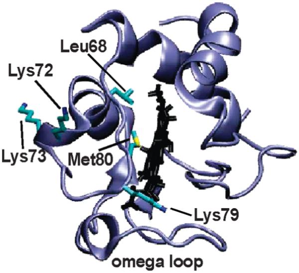
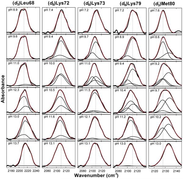
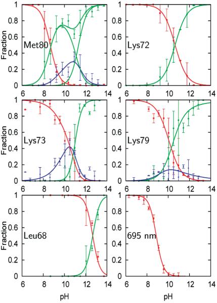
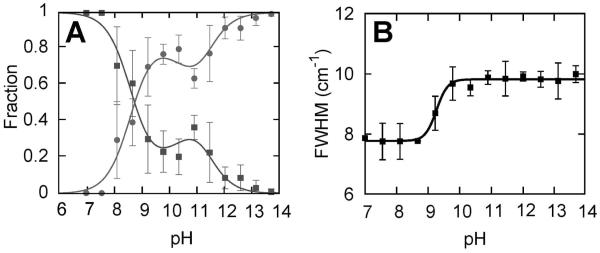
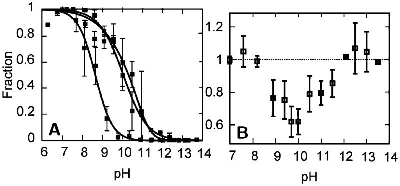
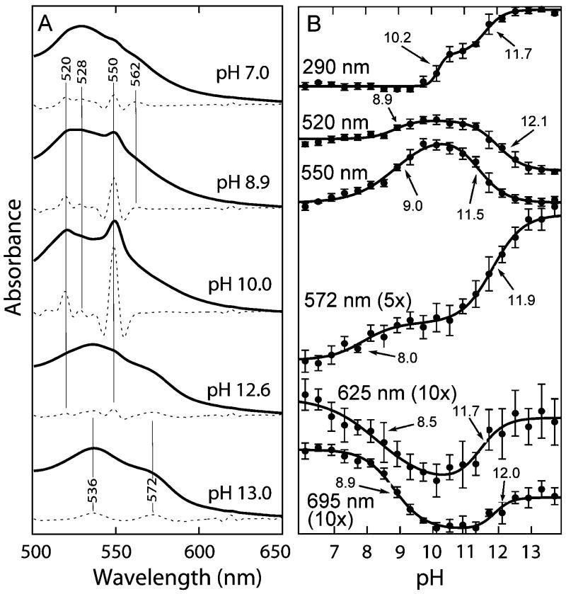
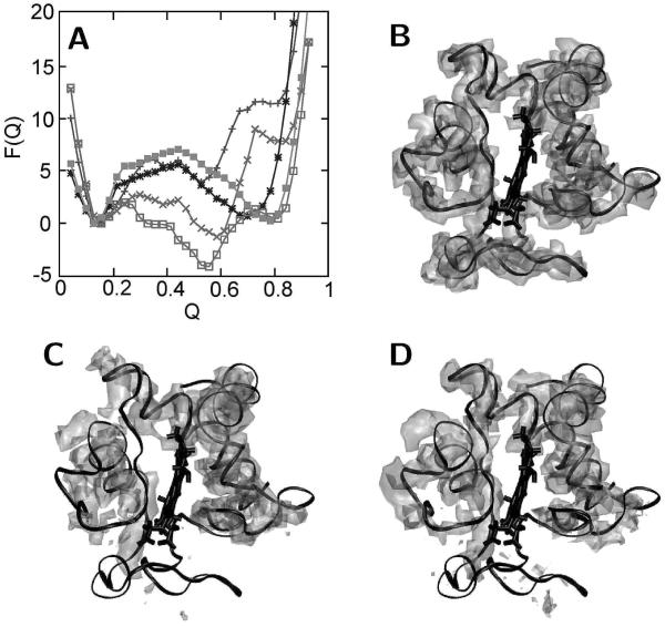
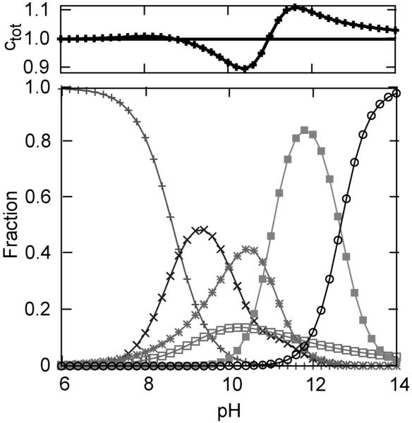
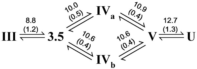
The alkaline-induced structural transitions of ferricytochrome c have been studied intensively as a model for how changes in metal ligation contribute to protein function and folding. Previous studies have demonstrated that multiple non-native species accumulate with increasing pH. Here, we used a combination of experiments and simulations to provide a high-resolution view of the changes associated with increasing alkaline conditions. Alkaline-induced transitions were characterized under equilibrium conditions by following changes in the IR absorptions of carbon-deuterium chromophores incorporated at Leu68, Lys72, Lys73, Lys79, and Met80. The data suggest that at least four intermediates are formed as the pH is increased prior to complete unfolding of the protein. The first alkaline transition observed appears to be driven by a single deprotonation and occurs with a midpoint of pH 8.8, but surprisingly, the intermediate formed does not appear to be one of the well-characterized lysine misligates. At higher pH, second and third deprotonations, with a combined apparent midpoint pH of 10.2, induce transitions to Lys73- or Lys79-misligated species. Interestingly, the lysine misligates appear to undergo iron reduction by the coordinated amine. A transition from the lysine misligates to another intermediate, likely a hydroxide-misligated species, is associated with a fourth deprotonation and a midpoint of pH 10.7. Finally, the protein loses tertiary structure with a fifth deprotonation that occurs with a midpoint of pH 12.7. Native topology-based models with enforced misligation are employed to help understand the structures of the observed intermediates.


Figures

Figure 1
Figure 1
Structure of cyt c (43) showing residues characterized.
Figure 2
Figure 2
Absorption spectra for each deuterated residue at several representative pH values (thick black lines). Also shown are the deconvolutions into folded, high pH/solvent-exposed, any intermediate signals (thin black lines), and resulting fits (red lines).
Figure 3
Figure 3
Fractional concentrations corresponding to folded (red), high pH, solvent-exposed (green), and any intermediate (blue) signals observed at each residue. Also shown are fractional concentrations derived from the intensity of the 695 nm absorption band (lower right panel). Data points (>60 per plot) have been binned into 0.5 pH intervals; error bars represent standard deviations. The curves obtained from model fitting are shown as lines (for details see text).
Figure 4
Figure 4
(A) Fractional concentrations corresponding to the folded and high pH, solvent-exposed signals resulting from a two-state fit of the (d3)Met80 data. (B) Line width (full width at half-maximum) of the (d3)Met80 folded signal resulting from a two-state fit in which the frequencies were fixed but the line widths were allowed to vary (for details see text).
Figure 5
Figure 5
(A) The fractional concentrations (also shown in Figure 3) of the folded signals for Met80 (lower pH transition) and Lys73/Lys79 (higher pH transitions). (B) Total fractional concentration for heme ligation by Met80, Lys73, Lys79, and lysine replacing ligand(s) calculated by summation of the fractional concentrations of the folded signal at Met80 and the intermediate signals and high pH, solvent-exposed signals at Lys73 and Lys79.
Figure 6
Figure 6
(A) UV/vis absorption spectra (solid lines) and their second derivatives (dashed lines) in the Q-band region at different pH values. (B) UV/vis absorption as a function of pH for several wavelengths. Midpoints are indicated with arrows and were obtained from three-state fits.
Figure 7
Figure 7
Results of the minimalist model simulations. (A) Free energy curves as a function of the reaction coordinate Q (related to the fraction of native contacts) obtained from simulation with enforced heme ligation of Lys53 (■), Lys55 (*), Lys72 (+), Lys73 (□), and Lys79 (×). (B–D) Isodensity surface plots determined using structures from the stable free energy basins calculated from simulations for (B) the native state, (C) the Lys73-misligated state, and (D) the Lys79-misligated state. The crystal structure of cyt c (black) is superimposed for comparison (see text for details).
Figure 8
Figure 8
Fractional concentrations derived by combining signals from Figure 3 to represent states III (+, native), 3.5 (×, neither methionine nor lysine ligated), IVa (*, Lys73 ligated), IVb(□, Lys79 ligated), V (■, hydroxide ligated), and U (○, unfolded). The total concentration (Ctot) obtained by summing over the fractional concentrations of the six states is shown in the upper box. See text for details.
Figure 9
Figure 9
Absorption spectra for states III, 3.5, IVa, IVb, V, and U determined by a deconvolution of the UV/vis data using the fractional concentrations determined from the IR data (see text and Supporting Information for details).
Scheme 1
Scheme 1
a a The observed transitions are shown with the apparent midpoint and n values (in parentheses) above each arrow.

References

    1. McLachlan AD. Rapid comparison of protein structures. Acta Crystallogr. 1982;A38:871–873.
    1. Weinkam P, Zong C, Wolynes PG. A funneled energy landscape for cytochrome c directly predicts the sequential folding route inferred from hydrogen exchange experiments. Proc. Natl. Acad. Sci. U.S.A. 2005;102:12401–12406. - PMC - PubMed
    1. Scott RA, Mauk AG. University Science Books; Sausalito, CA: 1996. pp. 611–634.
    1. Jones CM, Henry ER, Hu Y, Chan C, Luck SD, Bhuyan A, Roder H, Hofrichter J, Eaton WA. Fast events in protein folding initiated by nanosecond laser photolysis. Proc. Natl. Acad. Sci. U.S.A. 1993;90:11860–11864. - PMC - PubMed
    1. Theorell H, Akesson AJ. Studies on cytochrome c. II. The optical properties of pure cytochrome c and some of its derivatives. J. Am. Chem. Soc. 1941;63:1812–1818.

Publication types

Associated data