

Guideline
doi: 10.1111/j.1538-7836.2008.03242.x.
Epub 2008 Nov 25.
Recommendations for nomenclature on fibrinogen and fibrin
Affiliations
- PMID: 19036059
- PMCID: PMC3307547
- DOI: 10.1111/j.1538-7836.2008.03242.x
Item in Clipboard
Guideline
Recommendations for nomenclature on fibrinogen and fibrin
J Thromb Haemost.
2009 Feb.
No abstract available
Figures
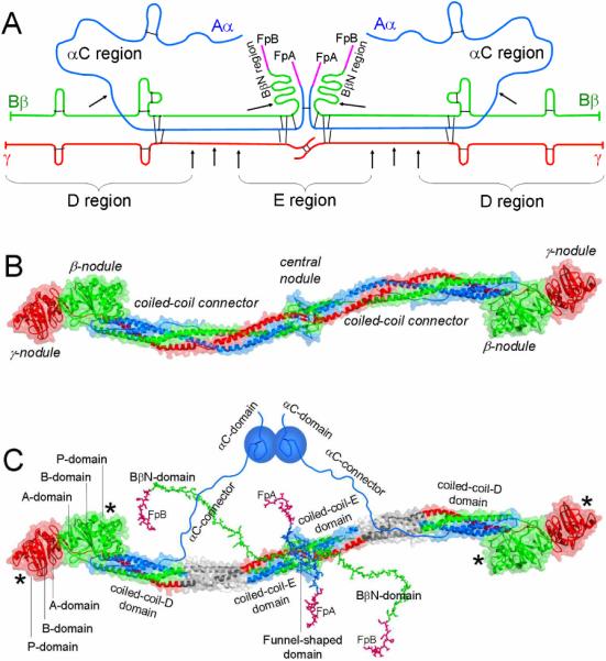
Panel A, polypeptide chain composition of fibrinogen. The individual chains, Aα, Bβ and γ, are blue, green and red, respectively; fibrinopeptides A and B (FpA and FpB) are magenta; the disulfide bonds are shown by black bars; triple arrows show proteolytic cleavages between the D and E regions, single arrows show cleavages resulting in the removal of the αC and BbN regions. Panel B, crystal structure of fibrinogen [8]. The central nodule is formed by the NH2-terminal portions of all six chains; it is connected to the distal β- and γ-nodules formed by the COOH-terminal portions of the Bβ and γ chains, respectively, by triple-helical coiled-coil connectors, each formed by the middle portions of the Aα, Bβ and γ chains. The color scheme is the same as in panel A. Panel C shows the same molecule as in panel B plus those regions that were not identified in the crystal structure, the interacting αC-domains, which are attached to the bulk of the molecule with the flexible αC-connectors, and the NH2-terminal portions of the Bβ chains forming the BbN regions (functional BβN-domains). Note, that the BβN-domains are shown in random conformation as in [23], and that this model does not present their interaction with the αC-domains identified in [24]. The funnel-shaped domain in the center contains fibrinopeptides A (colored magenta), which are also shown in random conformation as in [23]; the γN-domain [6] is located on the opposite side of the molecule and is not visible. The individual domains of the D regions, A-domain, B-domain, and P-domain, are indicated only in one subunit of the molecule. The site `a' or hole 'a' and site `b' or hole `b' in the P-domain of the γ- and β-nodules, respectively, are indicated by asterisks. The three chains are colored as in panels A and B, their protease sensitive portions between the D and E regions, which are not present in the D and E fragments, are shown in grey.
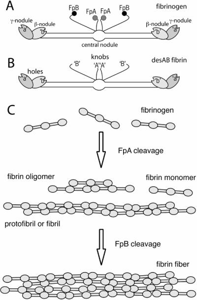
Schematic diagrams of fibrinogen (panel A), showing FpA, FpB, hole `a', hole `b', and desAB fibrin monomer (panel B), showing the exposure of knobs `A' with cleavage of FpA and the exposure of knobs `B' with cleavage of FpB. Panel C, initial steps of fibrin polymerization, showing fibrin oligomers, protofibrils or fibrils, and fibers.
References
-
- Henschen A, McDonagh J. Fibrinogen, fibrin and factor XIII. In: Zwaal RFA, Hemker HC, editors. Blood Coagulation. Elsevier Science Publishers; Amsterdam: 1986. pp. 171–241.
-
- Nussenzweig V, Seligmann M, Pelimont J, Grabar P. Les produits de degradation du fibrinogene humain par la plasmine. Ann Inst Pasteur. 1961;100:377–89. - PubMed
-
- Privalov PL, Medved LV. Domains in the fibrinogen molecule. J Mol Biol. 1982;159:665–83. - PubMed
-
- Spraggon G, Everse SJ, Doolittle RF. Crystal structures of fragment D from human fibrinogen and its crosslinked counterpart from fibrin. Nature. 1997;389:455–62. - PubMed
Publication types
MeSH terms
Substances
Grants and funding
LinkOut - more resources
Full Text Sources
