

Selective receptor expression restricts Nipah virus infection of endothelial cells
- PMID: 19036148
- PMCID: PMC2607271
- DOI: 10.1186/1743-422X-5-142
Selective receptor expression restricts Nipah virus infection of endothelial cells
Abstract
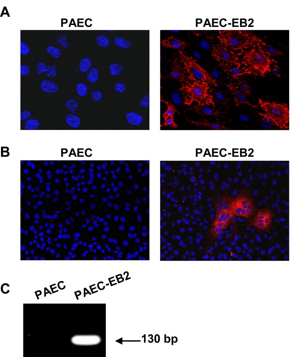
Nipah virus (NiV) is a highly pathogenic paramyxovirus that causes severe diseases in animals and humans. Endothelial cell (EC) infection is an established hallmark of NiV infection in vivo. Despite systemic virus spread via the vascular system, EC in brain and lung are preferentially infected whereas EC in other organs are less affected. As in vivo, we found differences in the infection of EC in cell culture. Only brain-derived primary or immortalized EC were found to be permissive to NiV infection. Using a replication-independent fusion assay, we could show that the lack of infection in non-brain EC was due to a lack of receptor expression. The NiV entry receptors ephrinB2 (EB2) or ephrinB3 were only expressed in brain endothelia. The finding that EB2 expression in previously non-permissive aortic EC rendered the cells permissive to infection then demonstrated that EB2 is not only necessary but also sufficient to allow the establishment of a productive NiV infection. This strongly suggests that limitations in receptor expression restrict virus entry in certain EC subsets in vivo, and are thus responsible for the differences in EC tropism observed in human and animal NiV infections.
Figures


Similar articles
-
Ephrin-B2 expression critically influences Nipah virus infection independent of its cytoplasmic tail.Virol J. 2008 Dec 24;5:163. doi: 10.1186/1743-422X-5-163. Virol J. 2008. PMID: 19108727 Free PMC article.
-
Two key residues in ephrinB3 are critical for its use as an alternative receptor for Nipah virus.PLoS Pathog. 2006 Feb;2(2):e7. doi: 10.1371/journal.ppat.0020007. Epub 2006 Feb 10. PLoS Pathog. 2006. PMID: 16477309 Free PMC article.
-
EphrinB2 is the entry receptor for Nipah virus, an emergent deadly paramyxovirus.Nature. 2005 Jul 21;436(7049):401-5. doi: 10.1038/nature03838. Epub 2005 Jul 6. Nature. 2005. PMID: 16007075
-
Henipavirus receptor usage and tropism.Curr Top Microbiol Immunol. 2012;359:59-78. doi: 10.1007/82_2012_222. Curr Top Microbiol Immunol. 2012. PMID: 22695915 Free PMC article. Review.
-
Envelope-receptor interactions in Nipah virus pathobiology.Ann N Y Acad Sci. 2007 Apr;1102(1):51-65. doi: 10.1196/annals.1408.004. Ann N Y Acad Sci. 2007. PMID: 17470911 Free PMC article. Review.
Cited by
-
The rising threat of Nipah virus: a highly contagious and deadly zoonotic pathogen.Virol J. 2025 May 10;22(1):139. doi: 10.1186/s12985-025-02728-4. Virol J. 2025. PMID: 40349023 Free PMC article. Review.
-
Generating human artery and vein cells from pluripotent stem cells highlights the arterial tropism of Nipah and Hendra viruses.Cell. 2022 Jul 7;185(14):2523-2541.e30. doi: 10.1016/j.cell.2022.05.024. Epub 2022 Jun 22. Cell. 2022. PMID: 35738284 Free PMC article.
-
Genome variations associated with viral susceptibility and calcification in Emiliania huxleyi.PLoS One. 2013 Nov 19;8(11):e80684. doi: 10.1371/journal.pone.0080684. eCollection 2013. PLoS One. 2013. PMID: 24260453 Free PMC article.
-
Characterization of a third generation lentiviral vector pseudotyped with Nipah virus envelope proteins for endothelial cell transduction.Gene Ther. 2013 Oct;20(10):997-1005. doi: 10.1038/gt.2013.23. Epub 2013 May 23. Gene Ther. 2013. PMID: 23698741 Free PMC article.
-
Henipavirus Immune Evasion and Pathogenesis Mechanisms: Lessons Learnt from Natural Infection and Animal Models.Viruses. 2022 Apr 29;14(5):936. doi: 10.3390/v14050936. Viruses. 2022. PMID: 35632678 Free PMC article. Review.
References
Publication types
MeSH terms
Substances
LinkOut - more resources
Full Text Sources
Research Materials
