. 2009 Mar 19;113(12):2835-42.
doi: 10.1182/blood-2008-06-159541. Epub 2008 Nov 25.

Platelets actively sequester angiogenesis regulators

Affiliations

Platelets actively sequester angiogenesis regulators

Giannoula Lakka Klement et al. Blood. .

Abstract

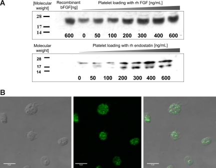
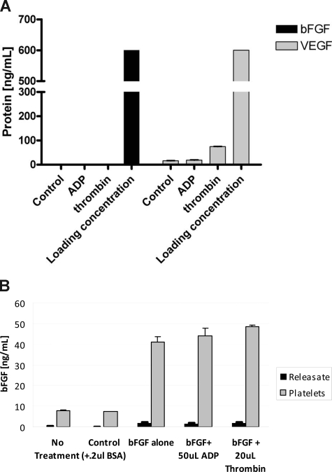
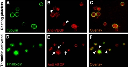
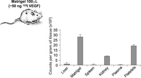
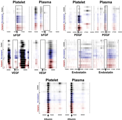
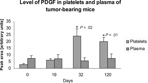
Clinical trials with antiangiogenic agents have not been able to validate plasma or serum levels of angiogenesis regulators as reliable markers of cancer presence or therapeutic response. We recently reported that platelets contain numerous proteins that regulate angiogenesis. We now show that accumulation of angiogenesis regulators in platelets of animals bearing malignant tumors exceeds significantly their concentration in plasma or serum, as well as their levels in platelets from non-tumor-bearing animals. This process is selective, as platelets do not take up a proportional amount of other plasma proteins (eg, albumin), even though these may be present at higher concentrations. We also find that VEGF-enriched Matrigel pellets implanted subcutaneously into mice or the minute quantities of VEGF secreted by microscopic subcutaneous tumors (0.5-1 mm(3)) result in an elevation of VEGF levels in platelets, without any changes in its plasma levels. The profile of other angiogenesis regulatory proteins (eg, platelet-derived growth factor, basic fibroblast growth factor) sequestered by platelets also reflects the presence of tumors in vivo before they can be macroscopically evident. The ability of platelets to selectively take up angiogenesis regulators in cancer-bearing hosts may have implications for the diagnosis and management of many angiogenesis-related diseases and provide a guide for antiangiogenic therapies.


Figures

Figure 1
Figure 1
Angiogenesis regulators are taken up by platelets in vitro. (A) PRP was incubated with increasing concentrations of recombinant human bFGF (rhbFGF) or recombinant human endostatin (rh endostatin) for an hour. The platelets were then isolated by sequential centrifugation, washed, treated with 1% Triton X to remove the membrane, and lysed for SDS-PAGE analysis. Standard Western blots using antihuman endostatin and antihuman bFGF antibodies reveal a dose-dependent increase in the respective proteins in the cytoplasmic fraction of fresh platelets. (B) To establish the localization of proteins taken up by platelets, the platelets were incubated with His-tag labeled endostatin, fixed using paraformaldehyde, and anti-His antibody was used to separate the platelet endogenous endostatin (not labeled) from the endostatin “loaded” into platelets (fluorescent label). (Left) DIC image of the platelets. (Middle) The fluorescent label of the His tag. (Right) The overlay of the 2 images. The pattern of the fluorescent signal indicates that endostatin is taken up into the granules of platelets rather than remaining membrane-associated.
Figure 2
Figure 2
Angiogenesis regulators loaded into platelets are not released with agonists of platelet activation. (A) A total of 1 mL PRP was incubated with 600 ng/mL of either VEGF or bFGF for 30 minutes. This resulted in “loading” of these proteins into the α-granules of platelets similarly to Figure 1. After the incubation, the platelets were aggregated using a mild aggregation agonist (ADP), a more potent agonist (thrombin), or spun down without any stimulation (control). The resulting supernatants/sera were then analyzed for VEGF and bFGF using a commercially available ELISA. Neither of the platelet agonists was capable of releasing bFGF from platelets. In the case of VEGF, ADP-induced aggregation failed to release the VEGF and even the more potent aggregation with thrombin resulted in only a modest release of the loaded VEGF. (B) PRP was incubated with 100 ng bFGF/mL on gentle rocker at room temperature for 45 minutes, spun at 150g, and the plasma containing excess of the protein was removed. The platelets were then resuspended in 1 mL saline to which 20 mM ADP or 1 unit thrombin was added. The sample was then spun again at 900g to pellet the platelets, and the supernatant and platelet analyzed using bFGF ELISA. As evident, platelets retained the majority of the loaded protein, and protein was released with either agonist. Both experiments were repeated on 2 separate occasions, and the graph represents the mean of 5 samples per dose level plus or minus SEM. Loading concentration in panel A corresponds to 600 ng VEGF or bFGF per mL.
Figure 3
Figure 3
VEGF localization in resting and activated platelets. Double-label immunofluorescence microscopy on fixed and permeabilized platelets was used to determine the intracellular localization of VEGF in the resting and activated states. In resting platelets, tubulin is concentrated, as expected, in the marginal microtubule band (A). In thrombin-activated platelets, phalloidin stain confirms the morphologic changes expected with activation (ie, the development of filopodia, lamellipodia, and pseudopodia) (D). The VEGF stain labels punctate, vesicle-like structures distributed throughout the platelet cytoplasm in both resting (arrowheads in panel B) and activated platelets (E), consistent with the lack of release of VEGF on activation, as noted in Figure 2. In the activated state, VEGF is redistributed to filopodia and lamellipodia (arrows in panel E and arrowheads in panel F) in contrast to its granular pattern of distribution in resting state (C).
Figure 4
Figure 4
Platelets selectively take up VEGF, without a corresponding increase of the protein in plasma. VEGF protein was labeled with radioactive iodine, and approximately 50 ng of 125I-labeled VEGF in 100 μL of Matrigel was implanted subcutaneously in the left flanks of C57/BL6 mice. Three days later, the mice were killed and 1 mL of citrated blood, as well as liver, kidney, spleen, and Matrigel pellet were collected. The radioactivity of each tissue sample was quantified on a gamma counter, the value corrected for differences in tissue weight, and expressed as counts per minute per gram of tissue. The experiment was repeated on 2 separate occasions with 5 mice per experiment, and the graph represents mean plus or minus SE of 1 of the experiments.
Figure 5
Figure 5
Platelet protein profiles of tumor-bearing mice correlate with tumor angiogenesis. SELDI-ToF MS expression difference maps from healthy mice (“Controls,” labeled gray), mice bearing nonangiogenic dormant tumor xenografts (“nonangiogenic,” labeled blue), and mice bearing angiogenic tumor xenografts of human liposarcoma (“angiogenic,” labeled red) are displayed in gel view. Differential expression patterns were detected for a range of peptides, and we present the most typical arrays: bFGF, PDGF, VEGF, endostatin, and albumin control. In each case, where differences in protein expressions were detected (eg, in the basic fraction [Q1 and Q2 fraction] of the platelet lysate, a difference was identified at 8200 Da), the band was further identified by purification and peptide mass fingerprinting and tandem MS (data not shown). (Abscissa) Relative MW computed from m/z value. (Ordinate) Identified peptide confirmed by immunodepletion and/or immunoprecipitation, and/or sequencing. The intensity of each of the bands correlates with relative expression level of the protein.
Figure 6
Figure 6
The tumor-associated change in platelet level of an angiogenesis regulator may be continuous. Platelet lysates from mice xenografted with human liposarcoma cells were longitudinally analyzed using SELDI-ToF at baseline, and up to 120 days after implantation of the tumor. The figure depicts an illustrative example of how the levels of an angiogenesis regulator (PDGF) are sustained in platelets for the duration of the experiment. Mice bearing dormant variant of human liposarcoma increase the platelet level of PDGF between 19 and 30 days after tumor implantation, and the level remains elevated for the duration of the experiment. The experiment was repeated on 2 separate occasions with 5 mice per experiment, and the graph represents mean plus or minus SEM.

Comment in

References

    1. Banks RE, Forbes MA, Kinsey SE, et al. Release of the angiogenic cytokine vascular endothelial growth factor (VEGF) from platelets: significance for VEGF measurements and cancer biology. Br J Cancer. 1998;77:956–964. - PMC - PubMed
    1. Kim HK, Song KS, Park YS, et al. Elevated levels of circulating platelet microparticles, VEGF, IL-6 and RANTES in patients with gastric cancer: possible role of a metastasis predictor. Eur J Cancer. 2003;39:184–191. - PubMed
    1. Karpatkin S. Tumor growth and metastasis. In: Michelson AD, editor. Platelets. San Diego, CA: Academic Press; 2002. pp. 491–502.
    1. Verheul HM, Pinedo HM. Tumor growth: a putative role for platelets? Oncologist. 1998;3:II. - PubMed
    1. Folkman J, Browder T, Palmblad J. Angiogenesis research: guidelines for translation to clinical application. Thromb Haemost. 2001;86:23–33. - PubMed

Publication types

MeSH terms