. 2009 Aug;50(8):1692-707.
doi: 10.1194/jlr.D800051-JLR200. Epub 2008 Nov 25.

Quantitative analysis of sphingolipids for lipidomics using triple quadrupole and quadrupole linear ion trap mass spectrometers

Affiliations

Quantitative analysis of sphingolipids for lipidomics using triple quadrupole and quadrupole linear ion trap mass spectrometers

Rebecca L Shaner et al. J Lipid Res. 2009 Aug.

Abstract

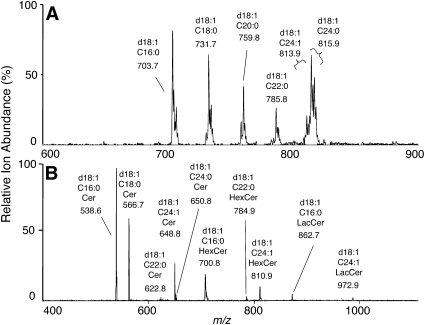
Sphingolipids are a highly diverse category of bioactive compounds. This article describes methods that have been validated for the extraction, liquid chromatographic (LC) separation, identification and quantitation of sphingolipids by electrospray ionization, tandem mass spectrometry (ESI-MS/MS) using triple quadrupole (QQQ, API 3000) and quadrupole-linear-ion trap (API 4000 QTrap, operating in QQQ mode) mass spectrometers. Advantages of the QTrap included: greater sensitivity, similar ionization efficiencies for sphingolipids with ceramide versus dihydroceramide backbones, and the ability to identify the ceramide backbone of sphingomyelins using a pseudo-MS3 protocol. Compounds that can be readily quantified using an internal standard cocktail developed by the LIPID MAPS Consortium are: sphingoid bases and sphingoid base 1-phosphates, more complex species such as ceramides, ceramide 1-phosphates, sphingomyelins, mono- and di-hexosylceramides, and these complex sphingolipids with dihydroceramide backbones. With minor modifications, glucosylceramides and galactosylceramides can be distinguished, and more complex species such as sulfatides can also be quantified, when the internal standards are available. LC ESI-MS/MS can be utilized to quantify a large number of structural and signaling sphingolipids using commercially available internal standards. The application of these methods is illustrated with RAW264.7 cells, a mouse macrophage cell line. These methods should be useful for a wide range of focused (sphingo)lipidomic investigations.


Figures

Fig. 1.
Fig. 1.
Workflow diagram for sample preparation for analysis of different categories of sphingolipids by LC-MS/MS. A: After addition of organic solvents to form a single phase and base hydrolysis (at far left), half of the sample is used as the “single-phase extract” for analysis of the shown categories of sphingolipids and the other half is further extracted to obtain a lower (organic) phase for the other categories. For more details, see “Materials and Methods.” B: Depiction of the major fragmentations of different categories of sphingolipids described in this article. For the sphingoid bases and ceramides depicted in the left two structures, -OR = -OH, -phosphate or mono- or di-hexosyl groups at position 1, and R and R′ are the alkyl sidechains for the sphingoid base and fatty acid, respectively, which can vary in length and, to some extent, unsaturation.
Fig. 2.
Fig. 2.
LC ESI-MS/MS elution profiles for the sphingolipids on reverse phase (A, B) and normal phase (C, D) chromatography. Shown are the elution of sphingoid bases and 1-phosphates and Cer1P in the single-phase extract of approximately 1 × 10⁶ RAW 264.7 cells with internal standard cocktail (500 pmol of each internal standard) (panel A) versus cells alone (panel B) from a Supelco 2.1mm i.d. × 5 cm Discovery C18 column and analysis by MRM in positive ionization mode as described under “Materials and Methods.” The abbreviations identify the nature of the sphingoid base (e.g., S, sphingosine, d18:1; Sa, sphinganine, d18:0; and internal standards d17:1 and d17:0), the 1-phosphates (1P), and ceramide1-phosphates (Cer1P, designating the sphingoid base and amide-linked fatty acid). C: Elution of complex sphingolipids in the “lower phase extract” of approximately 1 × 10⁶ RAW264.7 cells from a Supelco 2.1 mm i.d. × 5 cm LC-NH2 column and analysis by MRM in positive ion mode for Cer to LacCer, then negative ion mode for ST and Cer1P. D: Separation of d18:1/C16:0-GlcCer and -GalCer standards using a Supelco 2.1mm i.d. × 25 cm LC-Si column and the conditions described under “Materials and Methods.”
Fig. 3.
Fig. 3.
Product ion scans of sphinganine (A) and sphingosine (B) using the 4000 QTrap. Samples were infused in methanol.
Fig. 4.
Fig. 4.
Signal response for sphingoid bases and sphingoid base 1-phosphates using the ABI 3000 (QQQ) and 4000 QTrap. Each compound was analyzed over the shown range in amounts on column using the LC-MS/MS protocol for sphingoid bases and phosphates on reverse phase chromatography and positive ion mode MS/MS using optimized ionization, fragmentation, and MRM conditions as described under “Materials and Methods.”
Fig. 5.
Fig. 5.
Signal response for varying N-acyl chain length Cer (d18:1) and dihydroCer (d18:0) using the API 3000 (QQQ) and 4000 QTrap. Each compound was analyzed over the shown range in amounts on column using normal phase LC and positive ion mode MS/MS using optimized ionization, fragmentation, and MRM conditions as described under “Materials and Methods.”
Fig. 6.
Fig. 6.
Comparison of sphingomyelin product ion by traditional negative ion mode MS/MS in the 4000 QTrap (A) and by “pseudo MS3” analysis (B). Panel A is the product ion scan for the major precursor ion, m/z 631.5 from the in-source demethylation of SM; panel B reflects the results that are obtained by selecting the same precursor ion m/z for the first and second product ion, then offsetting Q2 by 5-10 eV so that no fragmentation occurs in the collision cell. Precursors are then induced to fragment in the ion trap by application of an amplitude frequency, allowing observation of the peaks shown when scanned as a “pseudo MS3.”
Fig. 7.
Fig. 7.
Precursor ion scans of RAW264.7 cells using the 4000 QTrap. The lower phase extract from approximately 106 RAW264.7 cells was infused in methanol and precursors ions that fragment to m/z 184.4 (panel A, sphingomyelins) and m/z 264.4 (panel B, Cer, HexCer and LacCer) are shown.
Fig. 8.
Fig. 8.
Recovery of internal standards and cellular sphingoid bases and 1-phosphates at each cycle of extraction. Approximately 1 × 10⁶ RAW264.7 cells (approximately 3 μ g DNA) were spiked with the internal standard cocktail (500 pmol each), extracted four times, and analyzed using LC-MS/MS on an 4000 QTrap mass spectrometer as described under “Materials and Methods.” The amounts of the analytes in each extract were calculated using the MRM areas for the unknowns versus the areas for the internal standards injected directly (i.e., without extraction). The bars represent the mean ± SD for n = 6.
Fig. 9.
Fig. 9.
Recovery of complex sphingolipids at each cycle of extraction. Approximately 1 × 10⁶ RAW264.7 cells (approximately 3 μ g DNA) were spiked with the internal standard cocktail (500 pmol of each species), extracted four times, and analyzed using LC-MS/MS on a 4000 QTrap mass spectrometer as described under “Materials and Methods.” The amounts of the analytes in each extract were calculated using the MRM areas for the unknowns versus the areas for the internal standards injected directly (i.e., without extraction). The bars represent the mean ± SD for n = 6.
Fig. 10.
Fig. 10.
Quantitative analysis of sphingolipids and dihydrosphingolipids in RAW264.7 cells. The amounts of these lipids were measured by LC-MS/MS using the internal standard cocktail as described under “Materials and Methods.” Shown are the means ± SE (n = 18) for three separate experiments with 6 dishes each. The upper insert shows the free sphingoid bases and 1-phosphates; the lower insert shows the amounts of sulfatides (ST) determined in an experiment where ST biosynthesis was induced by Kdo2-Lipid A, as described in the text. The N-acyl- chains of ST included hydroxy-24:1 (h24:1) and -24:0 (h24:0).

References

    1. Lahiri S., Futerman A. H. 2007. The metabolism and function of sphingolipids and glycosphingolipids. Cell. Mol. Life Sci. 64: 2270–2284 - PMC - PubMed
    1. Merrill A. H., Jr., Wang M. D., Park M., Sullards M. C. 2007. (Glyco)sphingolipidology: an amazing challenge and opportunity for systems biology. Trends Biochem. Sci. 32: 457–468 - PubMed
    1. van Echten-Deckert G.2000. Sphingolipid extraction and analysis by thin-layer chromatography. Methods Enzymol. 312: 64–79 - PubMed
    1. Muthing J.2000. Analyses of glycosphingolipids by high-performance liquid chromatography. Methods Enzymol. 312: 45–64 - PubMed
    1. Mano N., Oda Y., Yamada K., Asakawa N., Katayama K. 1997. Simultaneous quantitative determination method for sphingolipid metabolites by liquid chromatography/ionspray ionization tandem mass spectrometry. Anal. Biochem. 244: 291–300 - PubMed

Publication types

MeSH terms

LinkOut - more resources