. 2009 Jan 23;284(4):2397-408.
doi: 10.1074/jbc.M808704200. Epub 2008 Nov 26.

Eomesodermin requires transforming growth factor-beta/activin signaling and binds Smad2 to activate mesodermal genes

Affiliations

Eomesodermin requires transforming growth factor-beta/activin signaling and binds Smad2 to activate mesodermal genes

Paola Picozzi et al. J Biol Chem. .

Abstract

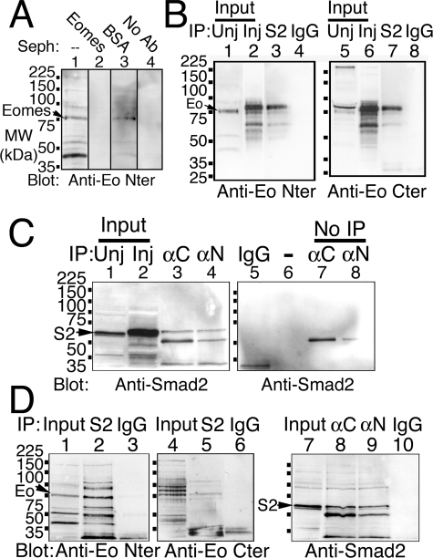
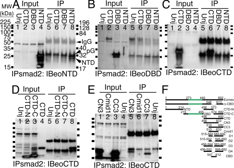
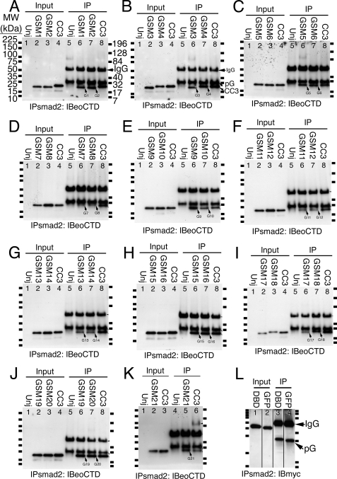
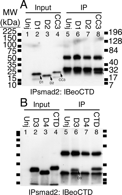
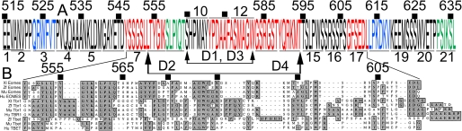
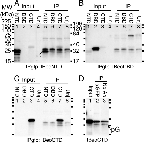
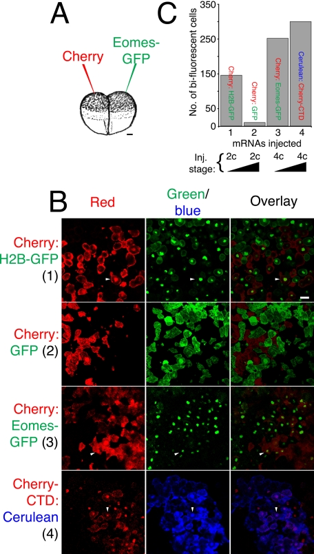
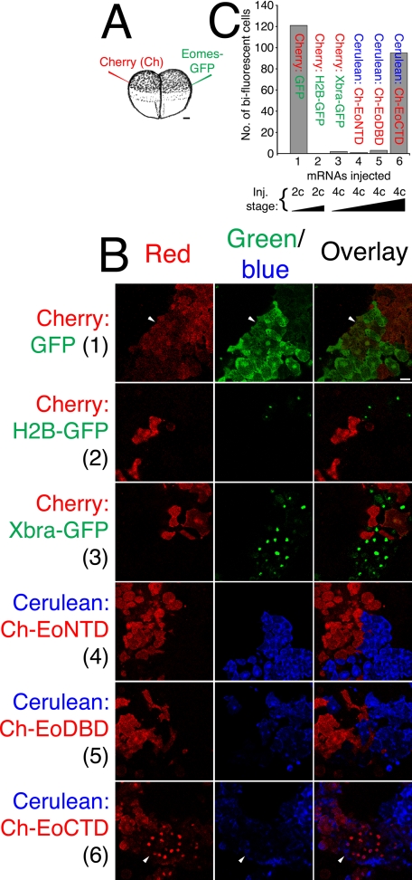
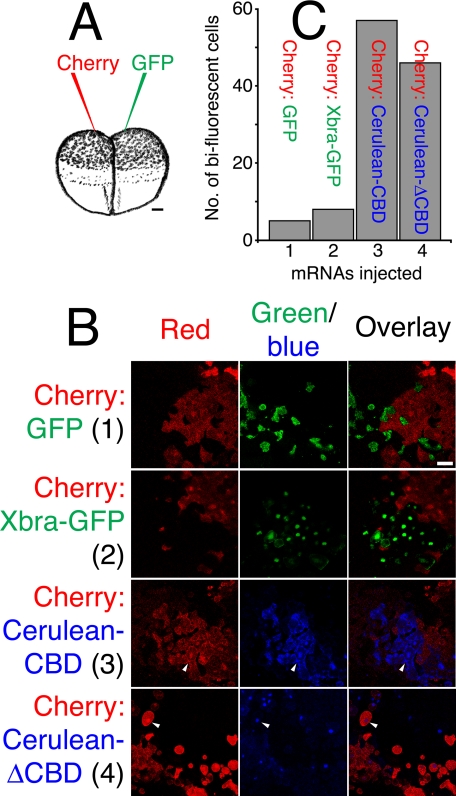
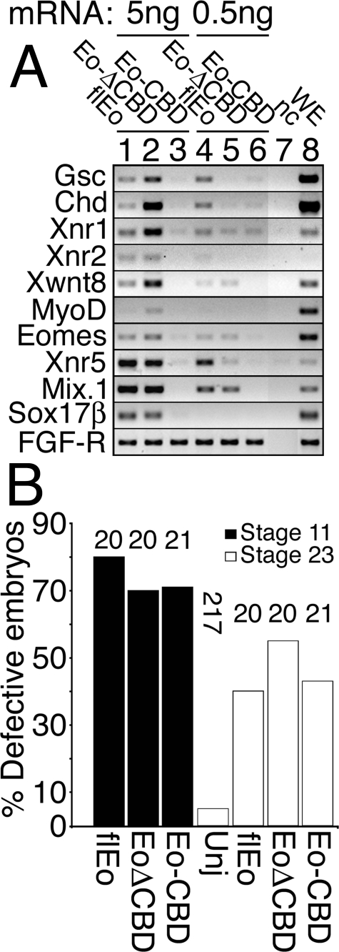
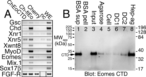
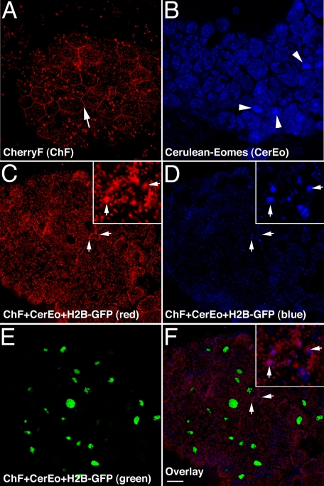
The T-box gene Eomesodermin (Eomes) is required for early embryonic mesoderm differentiation in mouse, frog (Xenopus laevis), and zebrafish, is important in late cardiac development in Xenopus, and for CD8+ T effector cell function in mouse. Eomes can ectopically activate many mesodermal genes. However, the mechanism by which Eomes activates transcription of these genes is poorly understood. We report that Eomes protein interacts with Smad2 and is capable of working in a non-cell autonomous manner via transfer of Eomes protein between adjacent embryonic cells. Blocking of Eomes protein transfer using a farnesylated red fluorescent protein (CherryF) also prevents Eomes nuclear accumulation. Transfer of Eomes protein between cells is mediated by the Eomes carboxyl terminus (456-692). A carbohydrate binding domain within the Eomes carboxyl-terminal region is sufficient for transfer and important for gene activation. We propose a novel mechanism by which Eomes helps effect a cellular response to a morphogen gradient.


Figures

FIGURE 1.
FIGURE 1.
Eomes protein interacts with Smad2. A, the anti-Eomes NH2-terminal antibody is specifically blocked by addition of the Eomes NH2-terminal antigen. Lysates from stage 10.5 embryos were left untreated (lanes 1 and 4), or immunodepleted by incubation with Sepharose that had been covalently cross-linked to the Eomes NH2-terminal antigen (lane 2) or bovine serum albumin (lane 3); immunodepleted lysates were Western blotted with the anti-Eomes NH2-terminal antibody (lanes 1-3) or no antibody (lane 4). B, overexpressed Eomes and Smad2 interact in vivo in Xenopus embryos. Lysates from uninjected stage 10.5 embryos (lanes 1, and 5), or embryos that had been injected with mRNAs for Eomes plus Smad2 (lanes 2 and 6), were immunoprecipitated with antibodies to Smad2 (lanes 3 and 7), or an irrelevant GFP antibody (lanes 4 and 8); immunoprecipitates were Western blotted with the Eomes NH2-terminal (lanes 1-4) or COOH-terminal (lanes 5-8) antibodies. C, overexpressed Smad2 and Eomes interact in vivo in Xenopus embryos. Smad2 and Eomes were coexpressed in Xenopus embryos. Uninjected extract (lane 1) or Eomes antibodies alone (lanes 7 and 8) are shown for comparison; co-injected extracts (lane 2) were immunoprecipitated with anti-Eomes antibodies (anti-COOH terminus, lane 3; anti-NH2 terminus, lane 4), or an irrelevant GFP antibody (lane 5); immunoprecipitates were Western blotted with an anti-Smad2 antibody. (No protein was loaded in lane 6.) D, endogenous Eomes and Smad2 interact in vivo in Xenopus embryos. Extracts (lanes 1, 4, and 7) were immunoprecipitated with anti-Smad2 antibody (lanes 2 and 5), anti-Eomes antibodies (lanes 8 and 9), or a GFP antibody (lanes 3, 6, and 10); immunoprecipitates were Western blotted using anti-Eomes antibodies (lanes 1-6) or anti-Smad2 antibody (lanes 7-10).
FIGURE 2.
FIGURE 2.
Thecentral region of the Eomes CTD interacts with Sepharose beads. A, the Eomes NTD largely failed to bind to Sepharose. The Eomes NTD, DBD, or CTD were overexpressed in Xenopus embryos. Extracts were left untreated (lanes 1-4) or immunoprecipitated (lanes 5-8) with an anti-Smad2 antibody and Western blotted with anti-Eomes NH2-terminal antibodies. Uninjected extracts are shown for comparison (Unj, lanes 1 and 5). (Central clearing of some input bands was remediated by reducing input.) B, the Eomes DBD did not bind to beads. The blot in A was re-probed with anti-Eomes DBD antibodies. C, the Eomes CTD binds to beads. The blot in A was re-probed with anti-Eomes CTD antibodies. D, the NH2- and COOH-terminal halves of CTD (CTD-N, CTD-C) failed to bind to beads. CTD-N, CTD-C, or CTD were overexpressed in Xenopus embryos. Extracts were left untreated (lanes 1-4) or immunoprecipitated (lanes 5-8) with an anti-Smad2 antibody and Western blotted with anti-Eomes COOH-terminal antibodies. E, both CN3 and CC3 bind to beads. CN3, Cmid1, or CC3 were overexpressed in Xenopus embryos. Extracts were left untreated (lanes 1-4) or immunoprecipitated (lanes 5-8) with an anti-Smad2 antibody and Western blotted with anti-Eomes COOH-terminal antibodies. (The anti-Eomes-CTD antibody failed to recognize Cmid1 (lanes 2 and 6)). F, Eomes constructs used in co-immunoprecipitation analysis. Eomes is 692 amino acids long. The carboxyl-terminal region amino acid residues 547-612 are deleted in Δ-CBD. NTD encodes 1-214; CTD-N, 456-573; DBD, 215-455; CTD-C, 574-692; CTD, 456-692; CN3, 456-633; CC3, 515-692; Cmid1, 515-633; D1, 515-564 fused to 583-692; D2, 515-552 fused to 595-692; D3, 456-564 fused to 583-692; D4, 456-552 fused to 595-692; CBD, 547-612. Eomes T-box (273-440) highlighted green.
FIGURE 3.
FIGURE 3.
The Eomes carboxyl-terminal region contains a carbohydrate binding domain. A, GSM1 and GSM2 bind to Sepharose beads. Eomes glycine-scanning mutant GSM1, GSM2, or CC3, were overexpressed in Xenopus embryos. Extracts were left untreated (lanes 1-4) or immunoprecipitated (lanes 5-8) with an anti-Smad2 antibody and Western blotted with anti-Eomes COOH-terminal antibodies. Uninjected extracts are shown for comparison (Unj, lanes 1 and 5). (GSM1, -2 lack a myc epitope tag.) B, GSM3 was slightly diminished, GSM4 binds to beads. (GSM3 lacks myc.) C, GSM5, 6 bind beads. D, GSM7, 8 failed to bind beads. E, GSM9 was slightly increased, GSM10 binds beads. F, GSM11, 12 failed to bind beads. G, GSM13, 14 failed to bind beads. H, GSM15, 16 bind beads. I, GSM17 failed to bind beads; GSM18 was diminished. GSM18 migrated slower than expected on gels. J, GSM19, 20 bind beads. K, GSM21 was slightly increased in binding to beads. L, myc-tagged DBD or GFP failed to bind to beads. Eomes DBD or GFP was overexpressed in Xenopus embryos. Extracts were left untreated (lanes 1 and 2) or immunoprecipitated (lanes 3 and 4) with an anti-Smad2 antibody and Western blotted with an anti-myc epitope antibody. (The myc tag caused anomalously slow gel migration of DBD and GFP.)
FIGURE 4.
FIGURE 4.
Internal deletions within CTD or CC3 failed to bind to Sepharose beads. A, D1 and D2 failed to bind to beads. Eomes deletion mutants D1, D2, or CC3 were overexpressed in Xenopus embryos. Extracts were left untreated (lanes 1-4) or immunoprecipitated (lanes 5-8) with an anti-Smad2 antibody and Western blotted with anti-Eomes COOH-terminal antibodies. Uninjected extracts are shown for comparison (Unj, lanes 1 and 5). B, deletion mutant D3 in CTD binds to beads; D4 failed to bind to beads.
FIGURE 5.
FIGURE 5.
The Eomes CBD is evolutionarily conserved. A, summary of results from glycine-scanning and deletion mutation of Eomes. Glu515-Leu636 is depicted in which 21 GSMs were made: red letters indicate that binding to Sepharose beads was eliminated within a 6-amino acid window; blue, diminished binding; green, increased binding. Deletions are indicated by arrows; GSMs over- or underlined; and amino acid numbers bulleted above the sequence. B, ClustalW alignment of Eomes Tyr547-Leu612 (CBD) with Tbr1 and Tbet from Xenopus, zebrafish, mouse, human (GenBank accession numbers: xlEomes, P79944; zfEomes NP_57175; muEomes, O54839; huEOMES, NP_005433; xtTbr1, NP_001072587; zfTbr1, NP_001108562; muTbr1, Q64336; huTBR1, NP_006584; zfTbet, XP_001338262; muTbet, AF241242; huTBET, NP_037483.1).
FIGURE 6.
FIGURE 6.
The Eomes CTD interacts with Sepharose beads. A, the Eomes NTD largely failed to bind to Sepharose. The Eomes NTD, DBD, or CTD were overexpressed in Xenopus embryos. Extracts were left untreated (lanes 1-4) or immunoprecipitated (lanes 5-8) with an anti-GFP antibody and Western blotted with anti-Eomes NH2-terminal antibodies. Uninjected extracts are shown for comparison (Unj, lanes 4 and 8). B, the Eomes DBD did not bind to beads. The blot in A was re-probed with anti-Eomes DBD antibodies. C, the Eomes CTD binds to beads. The blot in A was re-probed with anti-Eomes CTD antibodies. D, CTD binds beads without added antibody. The Eomes CTD was overexpressed in Xenopus embryos. Extracts were left untreated (lane 1) or immunoprecipitated with an anti-GFP antibody (lane 2) or no antibody (lane 3), and Western blotted with anti-Eomes COOH-terminal antibodies.
FIGURE 7.
FIGURE 7.
Xenopus embryos can be used to measure intercellular translocation of proteins. A, experimental design. Depiction of two-cell Xenopus embryos, which were injected into the animal pole. One cell was injected with mRNA encoding fluorescent protein of one color (e.g. Cherry), and the other cell with mRNA for another color (e.g. Eomes-GFP). Bar, 100 μm. B, mRNAs can diffuse between cells before completion of cytokinesis in two-cell embryos. One cell of mid-stage two-cell Xenopus embryos was injected with mRNA for Cherry, the other, histone H2B-GFP (top row). Animal poles were excised at stage 8 and subjected to confocal microscopy. Fluorescent images were obtained in the same confocal plane for each fluorescent color (shown individually and overlaid). Late two-cell embryos prevented diffusion of mRNA between cells (Cherry on one side, versus GFP on the other side; second row). Eomes protein is translocated between adjacent cells. Four-cell mid-stage embryos were injected with mRNA for Cherry on one side (left or right) versus Eomes-GFP on the other side (right or left; third row). The Eomes CTD is sufficient to mediate translocation of Eomes between cells. Late four-cell stage embryos were injected with mRNA for Cherry-Eomes-CTD on one side (left or right) versus Cerulean on the other side (right or left; fourth row). Bar, 38 μm. C, quantitation of Eomes protein translocation. The number of bi-fluorescent cells observed in 10 microscopic fields of view were counted. Views were randomly chosen and contained cells of both fluorescent colors. Arrowheads indicate bi-fluorescent cells.
FIGURE 8.
FIGURE 8.
Eomes intercellular translocation is mediated by the Eomes carboxyl-terminal region. A, two-cell Xenopus embryos were injected into the animal pole. One cell was injected with mRNA encoding fluorescent protein of one color, and the other cell with mRNA for another color. Injections were targeted at maximal distance from the embryonic midline (plane of first cell division). Bar, 100 μm. B, expression of Cherry in one cell and GFP in the other at the two-cell mid-stage allows diffusion of mRNA between cells (top row). Expression of Cherry versus H2B-GFP in late two-cell embryos prevents mRNA diffusion (second row). Xbra-GFP did not move between cells (versus Cherry, third row). Neither Cherry-Eomes-NTD (versus Cerulean, fourth row) nor Cherry-Eomes-DBD (versus Cerulean, fifth row) moved between cells. Cherry-Eomes-CTD did move between cells (versus Cerulean, bottom row). Bar, 38 μm. C, quantitation of Eomes protein translocation. The number of bi-fluorescent cells observed in 10 microscopic fields of view were counted. Views were randomly chosen and contained cells of both fluorescent colors. Arrowheads indicate bi-fluorescent cells.
FIGURE 9.
FIGURE 9.
The Eomes CBD is sufficient, but not necessary for translocation of Eomes protein between cells. A, experimental design. Injections were performed at the four-cell stage. Bar, 100 μm. B, Cherry and GFP mRNAs did not diffuse, nor did their proteins move between cells (top row). Xbra failed to move between cells (Cherry versus Xbra-GFP, second row). The Eomes CBD was sufficient to confer intercellular translocation (Cherry versus Cerulean CBD, third row). The Eomes CBD was not necessary for protein translocation (Cherry versus Cerulean-ΔCBD, bottom row). Bar, 38 μm. C, quantitation of Eomes protein translocation. Arrowheads indicate bi-fluorescent cells.
FIGURE 10.
FIGURE 10.
The Eomes CBD is important in gene activation. A, Eomes-CBD failed to activate early embryonic genes. Although Eomes-ΔCBD activated genes at high dose (5 ng of mRNA per cap), Eomes-ΔCBD was significantly impaired for gene activation at moderate dose (0.5 ng per cap). Xenopus embryos were injected into the animal pole with mRNA encoding full-length Eomes (lanes 1 and 4), Eomes-ΔCBD (lanes 2 and 5), and CBD (lanes 3 and 6). Injected or uninjected (nc, lane 7) animal poles were explanted (caps) at stage 8 and cultured until stage 10.5; and caps were frozen. Caps and control stage 10.5 whole embryos (WE, lane 8) were analyzed by RT-PCR for early embryonic genes. B, quantitation of embryonic defects in animal pole-injected, and uninjected, embryos. Embryos were scored for normalcy of gastrulation at stage 11, and for defects resulting from earlier inhibition of gastrulation at stage 23. Number of embryos (n) is shown above the histogram.
FIGURE 11.
FIGURE 11.
The Eomes CTD activates Xnr5 but fails to bind DNA. A, Cherry-Eomes-CTD activated Xnr5. Xenopus embryos were injected into the animal pole with mRNA encoding Cherry-Eomes-CTD, Cherry-f.l.Eomes (full-length), or Cherry alone; injected or uninjected (nc) animal poles were explanted (caps) at stage 8 and cultured until stage 10.5; and caps were frozen. Caps and control stage 10.5 whole embryos (WE) were analyzed by RT-PCR for early embryonic genes. B, the Eomes CTD failed to bind DNA. The Eomes CTD was overexpressed in Xenopus embryos. Extracts (lane 3) were co-precipitated with BSA cross-linked-agarose beads (lane 2), unsubstituted (plain) agarose (lane 4), DNase-treated DNA-cellulose (lane 5), mock-treated DNA-cellulose (lane 6), DNA-cellulose (lane 7), or heparin-agarose (lane 8), and Western blotted with anti-Eomes COOH-terminal antibodies. Supernatant from BSA beads is shown as a control (lane 1).
FIGURE 12.
FIGURE 12.
Eomes requires intercellular translocation for nuclear access. A, farnesylated Cherry (CherryF) binds to the plasma membrane (arrow). CherryF was expressed in caps and imaged by confocal microscopy. B, Cerulean-Eomes localizes to cell nuclei in caps (arrowheads). Cerulean-Eomes was expressed in caps and imaged by confocal microscopy. C-F were imaged in the same confocal plane; CherryF prevents Eomes accumulation in nuclei and pauses Cerulean-Eomes translocation at membrane puncta (arrows). CherryF, Cerulean-Eomes, and histone H2B-GFP were co-expressed in caps and imaged by confocal microscopy. Inset shows 4-fold magnification of membrane-localized puncta. C, red fluorescence; D, blue fluorescence; E, green fluorescence; F, overlay of C-E. Bar, 38 μm.

Similar articles

Cited by

References

    1. Ryan, K., and Chin, A. J. (2003) Birth Defects Res. Part C. Embryo Today 69 25-37 - PubMed
    1. Smith, J. (1999) Trends Genet. 15 154-158 - PubMed
    1. Conlon, F. L., Fairclough, L., Price, B. M., Casey, E. S., and Smith, J. C. (2001) Development 128 3749-3758 - PubMed
    1. Showell, C., Binder, O., and Conlon, F. L. (2004) Dev. Dyn. 229 201-218 - PMC - PubMed
    1. Wilson, V., and Conlon, F. L. (2002) Genome Biol. 3 REVIEWS3008 - PMC - PubMed

Publication types

MeSH terms

Associated data