. 2009 Feb 1;18(3):535-45.
doi: 10.1093/hmg/ddn381. Epub 2008 Nov 26.

Missense mutations that cause Van der Woude syndrome and popliteal pterygium syndrome affect the DNA-binding and transcriptional activation functions of IRF6

Affiliations

Missense mutations that cause Van der Woude syndrome and popliteal pterygium syndrome affect the DNA-binding and transcriptional activation functions of IRF6

Hayley J Little et al. Hum Mol Genet. .

Erratum in

  • Hum Mol Genet.2009 Apr 15;18(8):1544

Abstract

Cleft lip and cleft palate (CLP) are common disorders that occur either as part of a syndrome, where structures other than the lip and palate are affected, or in the absence of other anomalies. Van der Woude syndrome (VWS) and popliteal pterygium syndrome (PPS) are autosomal dominant disorders characterized by combinations of cleft lip, CLP, lip pits, skin-folds, syndactyly and oral adhesions which arise as the result of mutations in interferon regulatory factor 6 (IRF6). IRF6 belongs to a family of transcription factors that share a highly conserved N-terminal, DNA-binding domain and a less well-conserved protein-binding domain. To date, mutation analyses have suggested a broad genotype-phenotype correlation in which missense and nonsense mutations occurring throughout IRF6 may cause VWS; in contrast, PPS-causing mutations are highly associated with the DNA-binding domain, and appear to preferentially affect residues that are predicted to interact directly with the DNA. Nevertheless, this genotype-phenotype correlation is based on the analysis of structural models rather than on the investigation of the DNA-binding properties of IRF6. Moreover, the effects of mutations in the protein interaction domain have not been analysed. In the current investigation, we have determined the sequence to which IRF6 binds and used this sequence to analyse the effect of VWS- and PPS-associated mutations in the DNA-binding domain of IRF6. In addition, we have demonstrated that IRF6 functions as a co-operative transcriptional activator and that mutations in the protein interaction domain of IRF6 disrupt this activity.


Figures

Figure 1.
Figure 1.
IRF6 DNA-binding domain binds to ISRE sites. (A) SDS–PAGE gel showing purification of the IRF6 DNA-binding domain (IRF6-DBD). The protein was purified by Nickel-affinity chromatography. Fractions from each of the purification stages are shown as indicated above the lanes. Lane 5 contains the pure protein which was subsequently used to select the IRF6 consensus sequences. (B) EMSA showing DNA binding to the three pools of degenerate ISRE sequences as indicated above the lanes. The IRF6-DBD/DNA complexes are indicated by the arrow.
Figure 2.
Figure 2.
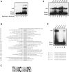
Selection of a consensus DNA-binding site for IRF6. (A) EMSA analysis of the selected pools of binding sites using bacterially expressed IRF6-DBD. The starting double-stranded DNA is shown in lane 0. The free DNA represents the DNA pool after the indicated number of rounds of selection. The position of the protein-DNA complex is shown (arrow). The DNA from the complexes in lane 3 was amplified and cloned for sequence analysis. (B) Sequences of the DNA-binding sites selected by IRF6 after three rounds of selection. Nucleotides derived from the random sequence (upper case) and the constant flanking primers (lower case) are indicated. The IRF core sequence is underlined in each sequence. Sites are aligned and orientated according to this IRF core sequence. (C) A schematic sequence representation for IRF6 binding sites after three rounds of selection. (D) EMSA showing IRF6-DBD binding to individual sequences obtained in the site selection. Lane 1 contains the initial sequence obtained from the IRF-E pool. The identity of each of the selected sites is shown above the lanes. (E) EMSA showing specific binding of IRF6-DBD to the S17 site and its variants containing specific point mutations. The identity of each of the sites is shown above the lanes. The core sequences are shown below the panel and the mutated bases are underlined.
Figure 3.
Figure 3.
DNA binding of VWS and PPS mutants. (A) The amino acid sequence of the IRF6 DNA-binding domain; a subset of mutations found in VWS and PPS patients are shown above the wild-type residue. (B) EMSA showing DNA binding of the mutant IRF6-DBD proteins depicted in (A). In vitro translated proteins were incubated with the consensus sequence shown above the panel. Lane 1 contains the wild-type IRF-DBD protein (WT). The identity of each mutant protein is indicated above the lanes; V18A, V18M, G70R, P76S, R84G, R84P, D98H and D98V are VWS-causing mutations: L22P and W60G are PPS-causing mutations; R84C, R84H and K89E underlie both VWS and PPS.
Figure 4.
Figure 4.
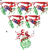
(A) Homology model of IRF6-DBD shown in red cartoon representation revealing a close-up of helix 3 after energy minimization The DNA is shown in green. Panel (i) shows the position of Arginine 84 in blue; panels (ii), (iii), (iv) and (v) show the mutations R84H, R84C, R84P and R84G, respectively. In each case, the Van der Waals surface around residue 84 is indicated with blue dots. (B) The position of the G70R mutation is shown, highlighting the distance of this residue from the DNA. Colour scheme as in (A).
Figure 5.
Figure 5.
Circular dichroism spectra of purified recombinant wild-type (WT) and mutant IRF6 proteins. The identity of each mutant protein is indicated in the key.
Figure 6.
Figure 6.
IRF6 activates transcription. (A) Wild-type IRF6 and a series of N-terminal deletions were fused to the C-terminus of the GAL4 DNA-binding domain as depicted. The DNA-binding and the protein interaction domains are indicated. The positions of the N- and C-terminal amino acids are shown for each of the IRF6 derivatives. (B) The graph shows activation of the luciferase reporter by the GAL-IRF6 proteins in the presence and absence of a fixed amount of LEXA-VP16 as indicated by + and −, respectively. The identity of the GAL fusion proteins in each transfection is indicated below the x-axis. All transfections were performed in triplicate; luciferase activities are presented as means with standard errors shown. All values are relative to the activity of the reporter plasmid alone. (C) Titration of increasing amounts of GAL-IRF6-(226-467). The graph shows activation of the luciferase reporter by the GAL-IRF6-(226-467) protein in the presence and absence of a fixed amount of LEXA-VP16 as indicated by + and −, respectively. 0, 10, 100 and 300 ng of GAL-IRF6-(226-467) were transfected. All transfections were performed in triplicate; luciferase activities are presented as means with standard errors shown. All values are relative to the activity of the reporter plasmid alone.
Figure 7.
Figure 7.
The effect of VWS and PPS mutations on the function of the IRF6 transactivation domain. (A and B) The graphs show activation of the luciferase reporter by the wild-type (WT) and mutant GAL-IRF6-(226-467) proteins in the presence and absence of a fixed amount of LEXA-VP16 as indicated by + and −, respectively. The identity of each mutation is indicated below the x-axis. All transfections were performed in triplicate; luciferase activities are presented as means with standard errors shown. All values are relative to the activity of the reporter plasmid alone.

References

    1. Vanderas A.P. Incidence of cleft lip, cleft palate, and cleft lip and palate among races: a review. Cleft Palate J. 1987;24:216–225. - PubMed
    1. Murray J.C., Daack-Hirsch S., Buetow K.H., Munger R., Espina L., Paglinawan N., Villanueva E., Rary J., Magee K., Magee W. Clinical and epidemiologic studies of cleft lip and palate in the Philippines. Cleft Palate Craniofac. J. 1997;34:7–10. - PubMed
    1. Neilson D.E., Brunger J.W., Heeger S., Bamshad M., Robin N.H. Mixed clefting type in Rapp-Hodgkin syndrome. Am. J. Med. Genet. 2002;108:281–284. - PubMed
    1. Jones M.C. Etiology of facial clefts: prospective evaluation of 428 patients. Cleft Palate J. 1988;25:16–20. - PubMed
    1. Suzuki K., Hu D., Bustos T., Zlotogora J., Richieri-Costa A., Helms J.A., Spritz R.A. Mutations of PVRL1, encoding a cell-cell adhesion molecule/herpesvirus receptor, in cleft lip/palate-ectodermal dysplasia. Nat. Genet. 2000;25:427–430. - PubMed

Publication types

MeSH terms

Substances