. 2009 Feb;200(2):207-21.
doi: 10.1677/JOE-08-0241. Epub 2008 Nov 26.

Vitamin D reduces the expression of collagen and key profibrotic factors by inducing an antifibrotic phenotype in mesenchymal multipotent cells

Affiliations

Vitamin D reduces the expression of collagen and key profibrotic factors by inducing an antifibrotic phenotype in mesenchymal multipotent cells

Jorge N Artaza et al. J Endocrinol. 2009 Feb.

Abstract

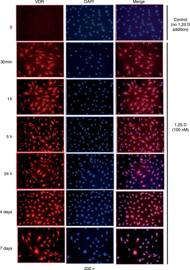
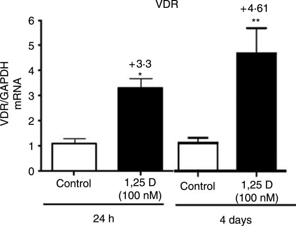
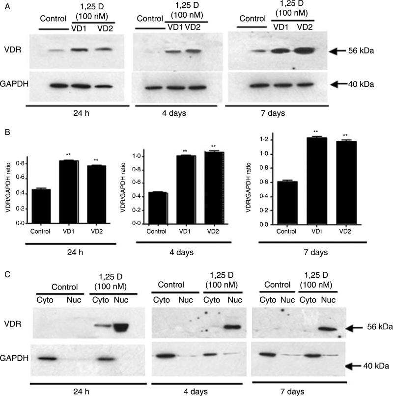
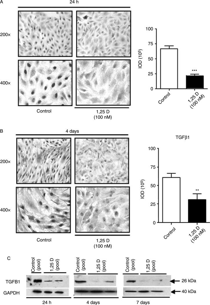
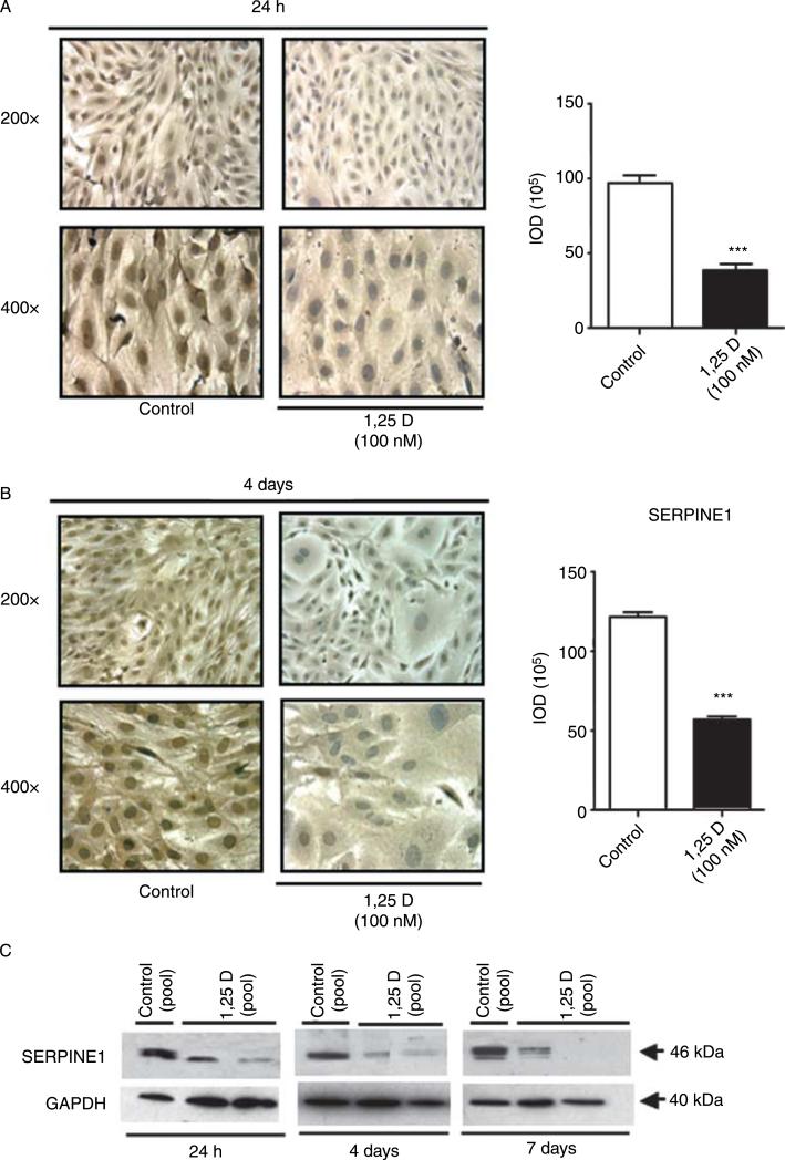
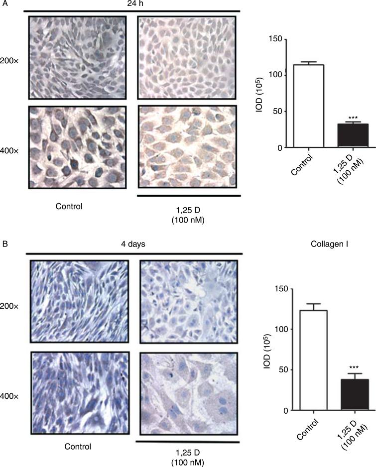
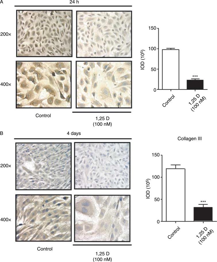
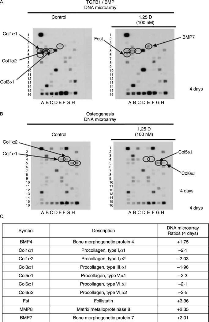
Hypovitaminosis D is an important public health problem. Serum 25-hydroxyvitamin D (25-OHD) is now recognized as an independent predictor for cardiovascular and related diseases (CVD) as well as other chronic medical conditions. However, the biologic pathways through which these effects are mediated remain poorly understood. We hypothesized that exposing mesenchymal multipotent cells (MMCs) to the active form of vitamin D would increase the expression of selected antifibrotic factors that in turn would ameliorate the progression of chronic diseases. MMCs were primed with 5'-azacytidine to induce a fibrotic phenotype and then treated with active vitamin D (1,25D) or ethanol <0.1% as vehicle in a time course manner (30 min, 1, 5, and 24 h, and for 4 and 7 days). The addition of 1,25D to MMCs promotes: a) increased expression and nuclear translocation of the vitamin D receptor; b) decreased expression of TGFB1 and plasminogen activator inhibitor (SERPINE1), two well-known profibrotic factors; c) decreased expression of collagen I, III and other collagens isoforms; and d) increased expression of several antifibrotic factors such as BMP7 a TGFB1 antagonist, MMP8 a collagen breakdown inducer and follistatin, an inhibitor of the profibrotic factor myostatin. In conclusion, the addition of 1,25D to differentiated MMCs displays a decreased profibrotic signaling pathway and gene expression, leading to decrease in collagen deposition. This study highlights key mechanistic pathways through which vitamin D decreases fibrosis, and provides a rationale for studies to test vitamin D supplementation as a preventive and/or early treatment strategy for CVD and related fibrotic disorders.


Figures

Figure 1
Figure 1
Time course of the expression and nuclear translocation of vitamin D receptor (VDR) upon incubation with 1,25D in mesenchymal multipotent C3H 10T1/2 cells. Cultures of C3H 10T1/2 cells were treated with 20 μM azacytidine for 2 days to induce fibrosis and 2 days later were incubated with or without 1,25D (100 nM) at different time points: 30 min, 1, 5, and 24 h, and for 4 and 7 days. Cells were cultured on eight-well removable chambers slides and subjected for immunofluorescence using a polyclonal antibody for VDR followed by a Texas Red-conjugated secondary antibody (red). Cells were counterstained with DAPI (blue) to show nuclear staining. Merge pictures were done combining the red and blue pictures together (purple) and in order to show nuclear localization of VDR. Magnification=200×.
Figure 2
Figure 2
Transcriptional upregulation of mRNA VDR upon incubation with 1,25D in mesenchymal multipotent C3H 10T1/2 cells. Cultures of C3H 10T1/2 cells were treated as in Fig. 1 and incubated with or without 1,25D (100 nM) for 24 h and 4 days. Total RNA isolation followed by real-time RT-PCR was applied and normalized by GAPDH housekeeping gene. Mean±s.e.m.. corresponds to experiments done in triplicates. *P<0·05, **P<0·01.
Figure 3
Figure 3
Translational upregulation of VDR expression and nuclear translocation upon incubation with 1,25D in mesenchymal multipotent C3H 10T1/2 cells. Cultures of C3H 10T1/2 cells were treated as in Fig. 1 and incubated with or without 1,25D (100 nM) for 24 h, 4 and 7 days. Western blot immunoanalysis was performed in the whole extracts for each time point (A, top panel) and the corresponding quantitative densitometry analysis, **P<0·01 (A, bottom panel). Distribution of VDR protein in subcellular fractions (B) The nuclear and cytoplasmic fractions were isolated and subjected to western immunoblot analysis for VDR as described in the text. The same membrane in each case was stripped and reprobed with anti-GAPDH antibody to verify the purity of the subcellular fractionation.
Figure 4
Figure 4
Effects of 1,25D on the expression of the profibrotic marker TGFB1 in C3H 10T1/2 cells. Cultures of C3H 10T1/2 cells were treated as in Fig. 1 and incubated with or without 1,25D (100 nM) for 24 h, 4 and 7 days. Immunocytochemistry for TGFB1 were performed at 24 h (A) and 4 days (B) on eight-well removable chambers slides. Mean±s.e.m.. corresponds to experiments done in triplicates of the integrated optical density (IOD) by quantitative image analysis. **P<0·01, ***P<0·001. (C) Western blot analysis were performed with an anti-TGFB1 antibody to confirm the results obtained at 24 h, 4 and 7 days using a pool of cells done in triplicates from two different experiments. The same membrane was stripped and reprobed with anti-GAPDH antibody (housekeeping gene).
Figure 5
Figure 5
Effects of 1,25D on the expression of the profibrotic marker SERPINE1 in C3H 10T1/2 cells. Cultures of C3H 10T1/2 cells were treated as in Fig. 1 and incubated with or without 1,25D (100 nM) for 24 h, 4 and 7 days. Immunocytochemistry for SERPINE1 were performed at 24 h (A) and 4 days (B) on eight-well removable chambers slides. Mean±s.e.m.. corresponds to experiments done in triplicates of the integrated optical density (IOD) by quantitative imageanalysis ***P<0·001.(C) Western blot analysiswere performedwith an anti-SERPINE1 antibody to confirm the results obtained at 24 h, 4 and 7 days using a pool of cells done in triplicates from two different experiments. The same membrane was stripped and reprobed with anti-GAPDH antibody (housekeeping gene).
Figure 6
Figure 6
Effects of 1,25D on the expression of collagen I in C3H 10T1/2 cells. Cultures of C3H 10T1/2 cells were treated as in Fig. 1 and incubated with or without 1,25D (100 nM) for 24 h and 4 days. Immunocytochemistry for collagen I were performed at 24 h (A) and 4 days (B) on eight-well removable chambers slides. Mean±s.e.m.. corresponds to experiments done in triplicates of the integrated optical density (IOD) by quantitative image analysis ***P<0·001.
Figure 7
Figure 7
Effects of 1,25D on the expression of collagen III in C3H 10T1/2 cells. Cultures of C3H 10T1/2 cells were treated as in Fig. 1 and incubated with or without 1,25D (100 nM) for 24 h and 4 days. Immunocytochemistry for collagen III were performed at 24 h (A) and 4 days (B) on eight-well removable chambers slides. Mean±s.e.m.. corresponds to experiments done in triplicates of the integrated optical density (IOD) by quantitative image analysis ***P<0·001.
Figure 8
Figure 8
Effects of 1,25D on the transcriptional expression of different collagen isoforms and other fibrotic genes in C3H 10T1/2 cells by DNA microarrays. Cultures of C3H 10T1/2 cells were treated as in Fig. 1 and incubated with or without 1,25D (100 nM) for 4 days on 75 cm2 flasks. Total RNA was isolated and subjected to DNA microarray analysis for genes related to the TGFB1/BMP signaling pathway (A) and for osteogenesis gene array (B). Representative membranes for each array are displayed. The circles show changes in the expression of some antifibrotic and profibrotic genes that were upregulated or downregulated by 1,25D treatment. (C) Ratios between the 1,25D treated and 1,25D-untreated cells at 4 days, corrected by GAPDH were calculated for the assays performed in duplicates.

References

    1. Adams CD, Szumita PM, Baroletti SA, Lilly CM. Azacitidine-induced interstitial and alveolar fibrosis in a patient with myelodysplastic syndrome. Pharmacotherapy. 2005;25:765–768. - PubMed
    1. Amthor H, Nicholas G, McKinnell I, Kemp CF, Sharma M, Kambadur R, Patel K. Follistatin complexes myostatin and antagonises myostatin-mediated inhibition of myogenesis. Developmental Biology. 2004;270:19–30. - PubMed
    1. Aoki F, Kurabayashi M, Hasegawa Y, Kojima I. Attenuation of bleomycin-induced pulmonary fibrosis by follistatin. American Journal of Respiratory and Critical Care Medicine. 2005;172:713–720. - PubMed
    1. Artaza JN, Bhasin S, Mallidis C, Taylor W, Ma K, Gonzalez-Cadavid NF. Endogenous expression and localization of myostatin and its relation to myosin heavy chain distribution in C2C12 skeletal muscle cells. Journal of Cellular Physiology. 2002;190:170–179. - PubMed
    1. Artaza JN, Bhasin S, Magee TR, Reisz-Porszasz S, Shen R, Groome NP, Meerasahib MF, Gonzalez-Cadavid NF. Myostatin inhibits myogenesis and promotes adipogenesis in C3H 10T(1/2) mesenchymal multipotent cells. Endocrinology. 2005;146:3547–3557. - PubMed

Publication types

MeSH terms