. 2009 Jan;37(1):256-67.
doi: 10.1093/nar/gkn959. Epub 2008 Nov 26.

Function and ribosomal localization of aIF6, a translational regulator shared by archaea and eukarya

Affiliations

Function and ribosomal localization of aIF6, a translational regulator shared by archaea and eukarya

Dario Benelli et al. Nucleic Acids Res. 2009 Jan.

Abstract

The translation factor IF6 is shared by the Archaea and the Eukarya, but is not found in Bacteria. The properties of eukaryal IF6 (eIF6) have been extensively studied, but remain somewhat elusive. eIF6 behaves as a ribosome-anti-association factor and is involved in miRNA-mediated gene silencing; however, it also seems to participate in ribosome synthesis and export. Here we have determined the function and ribosomal localization of the archaeal (Sulfolobus solfataricus) IF6 homologue (aIF6). We find that aIF6 binds specifically to the 50S ribosomal subunits, hindering the formation of 70S ribosomes and strongly inhibiting translation. aIF6 is uniformly expressed along the cell cycle, but it is upregulated following both cold- and heat shock. The aIF6 ribosomal binding site lies in the middle of the 30-S interacting surface of the 50S subunit, including a number of critical RNA and protein determinants involved in subunit association. The data suggest that the IF6 protein evolved in the archaeal-eukaryal lineage to modulate translational efficiency under unfavourable environmental conditions, perhaps acquiring additional functions during eukaryotic evolution.


Figures

Figure 1.
Figure 1.
Sulfolobus solfataricus aIF6 interacts specifically with the 50S subunits. (a) Density gradient fractionation of crude, non-incubated, cell lysates. (b) Density gradient fractionation, after fixation with HCHO, of cell lysates programmed for translation and incubated at 70°C for 30 min. The distribution of aIF6, shown at the bottom of each gradient profile, was revealed by western blotting of the individual fractions with the anti-aIF6 antibodies. The distribution of the control protein L7ae, which is localized both in the cytoplasm and on the 50S ribosomal subunit, is also shown.
Figure 2.
Figure 2.
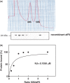
aIF6 has a single binding site on the 50S subunits. (a) Sucrose gradient fractionation of a preparation of S. solfataricus ribosomes incubated for 10 min at 65°C with an excess of recombinant aIF6. The presence of a IF6 in the gradient fractions was revealed by western blotting with the specific antibodies. (b) Binding curve of aIF6 to 50S ribosomes, showing saturation at a 1:1 protein/50S ratio. The amount of ribosome-bound aIF6 was evaluated as described in ‘Materials and methods’ section.
Figure 3.
Figure 3.
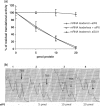
aIF6 blocks translation by inhibiting ribosomal subunit association. (a) Progressive inhibition of the translation of two reporter mRNAs, one leadered and one leaderless, upon addition of increasing amounts of recombinant aIF6 to a protein-synthesizing cell-free system. Lack of inhibition by the control protein aSUI1 is also shown. Each experimental point represents the average of three replicate experiments. (b) Density gradient fractionation of translation mixtures containing increasing amounts of recombinant aIF6 as indicated.
Figure 4.
Figure 4.
Expression of aIF6 in the cell cycle and upon thermal stress. Left: S. solfataricus cell cultures were grown to the indicated OD660; the cell lysates were electrophoresed and probed with the anti-aIF6 antibodies. Three replicate experiments were averaged, and the data were normalized using protein L7ae as the reference. Right: cells grown to the late exponential phase at 80°C were transferred for 30 min at 65°C for cold-shock and at 90°C for heat-shock; a control aliquot was left untreated. The amount of aIF6 in the cell lysates was evaluated by western blotting with the anti-aIF6 antibody and normalized using L7ae as the reference. The numbers on the vertical axis represent the fold increase in aIF6 amount relative to the 80°C value; each value represents the average of three replicate experiments.
Figure 5.
Figure 5.
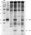
aIF6 interacts with ribosomal protein L14. Sypro-ruby-stained SDS–PAGE showing the protein bands recovered after immunoprecipitation of whole cell lysates or purified 50S subunits with the anti-aIF6 antibodies. Lane 1, control with recombinant aIF6; lanes 2 and 3, whole-cell lysates treated, respectively, with pre-immune serum or anti-aIF6 antibodies; lanes 4 and 5, 50S subunits treated, respectively, with pre-immune serum or anti-aIF6 antibodies. The bands containing aIF6 and L14 (indicated by the arrows) were unambiguously identified by MALDI-TOF/TOF.
Figure 6.
Figure 6.
Recombinant aIF6 and aL14 interact specifically in vitro. (a) Recombinant aIF6 and aL14 were incubated, separately and together, as described in the methods; the samples were treated with the anti-aIF6 antibodies or the correspondent pre-immune serum, electrophoresed and immunostained with the anti-His antibodies. Lanes 1 and 2, supernatant and precipitate, respectively, of a sample containing aIF6 treated with pre-immune serum; lanes 3 and 4, supernatant and precipitate, respectively, of samples containing aL14 treated with anti-aIF6 antibodies; lanes 5 and 6, supernatant and precipitate, respectively, of samples containing both aL14 and aIF6 treated with the anti-aIF6 antibodies. (b) Immunoprecipitation with the anti-aIF6 antibodies of samples containing aIF6 and the control ribosomal protein L7ae. Lanes 1 and 2, supernatant and precipitate, respectively, of samples containing aIF6 treated with the pre-immune serum; lanes 3 and 4, supernatant and precipitate, respectively, of samples containing aIF6 and L7ae treated with the anti-aIF6 antibodies. Staining was with the anti-His antibodies. HC: antibody heavy chain.
Figure 7.
Figure 7.
Identification of the 23S rRNA bases protected by bound aIF6. The left panel displays the autoradiography of a sequencing gel including samples of untreated 50S subunits (nm) and kethoxal-modified 50S subunit in the absence (-aIF6) or in the presence (+aIF6) of the recombinant aIF6 protein. A,C,G,U lanes show the sequence of the relevant rRNA region. The nucleotides significantly protected against modification by bound aIF6 are indicated by arrows; the reference band used for normalization is indicated by an asterisk. The primer used was complementary to region 2178–2198 of S. solfataricus 23S rRNA. The right panel shows a diagram of the secondary structure of S.solfataricus 23S rRNA domain IV (28), where the modified guanines are indicated by arrows.
Figure 8.
Figure 8.
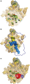
H69 modelling and docking of aIF6 on H. marismortui 50S. Cartoon and surface representations of Haloarcula marismortui 50S structure. (a, left) H69, in red, has been modeled using the structure of prokaryotic H69 (taken from the file 2I2V.pdb2) (19) using the programs Assemble and Pymol. (a, right) Nucleotides surrounding H69 protected by aIF6 are indicated by arrows; L14 is represented in dark blue solid surface. (b, left) H69, in red. aIF6 (solid red surface) has been docked using the constraints imposed by the protection of the 23S nucleotides and by the binding with L14 (blue solid surface). (b, right) Same, but rotated 80° around the Y axe.
Figure 9.
Figure 9.
Structural details of aIF6 anti-association function. Cartoon and surface representations of H. marismortui 50S structure as in Figure 7. (a) Intersubunit bridges anchoring the 30S subunit to the 50S. H69, in red. Green and cyan surface representations are 50S nucleotides and amino acids, respectively, involved in bridging the 30S subunit. (b) Representation of docked 30S Initiation Complex. Thermus thermophilus 70S X-ray structure (35) has been used to position the silhouette of the 30S subunit (blue profile), h44 (blue solid surface) and the initiator tRNA (yellow solid surface). (C) aIF6 prevent 70S association. The docking of aIF6 on the H. marismortui 50S prevent the formation of the most important bridges between the two subunits.

References

    1. Russell DW, Spremulli LL. Purification and characterization of a ribosome dissociation factor (eukaryotic initiation factor 6) from wheat germ. J. Biol. Chem. 1979;254:8796–8800. - PubMed
    1. Valenzuela DM, Chaudhuri A, Maitra U. Eukaryotic ribosomal subunit anti-association activity of calf liver is contained in a single polypeptide chain protein of Mr = 25,500 (eukaryotic initiation factor 6) J. Biol. Chem. 1982;257:7712–7719. - PubMed
    1. Basu U, Si K, Warner JR, Maitra U. The Saccharomyces cerevisiae TIF6 gene encoding translation initiation factor 6 is required for 60S ribosomal subunit biogenesis. Mol. Cell Biol. 2001;21:1453–1462. - PMC - PubMed
    1. Si K, Maitra U. The Saccharomyces cerevisiae homologue of mammalian translation initiation factor 6 does not function as a translation initiation factor. Mol. Cell Biol. 1999;19:1416–1426. - PMC - PubMed
    1. Basu U, Si K, Deng H, Maitra U. Phosphorylation of mammalian eukaryotic translation initiation factor 6 and its Saccharomyces cerevisiae homologue Tif6p: evidence that phosphorylation of Tif6p regulates its nucleocytoplasmic distribution and is required for yeast cell growth. Mol. Cell Biol. 2003;23:6187–6199. - PMC - PubMed

Publication types

MeSH terms