. 2009 Jan;37(1):243-55.
doi: 10.1093/nar/gkn938. Epub 2008 Nov 26.

Functional and structural characterization of the integrase from the prototype foamy virus

Affiliations

Functional and structural characterization of the integrase from the prototype foamy virus

Eugene Valkov et al. Nucleic Acids Res. 2009 Jan.

Abstract

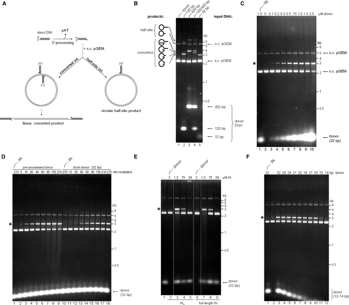
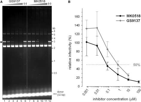
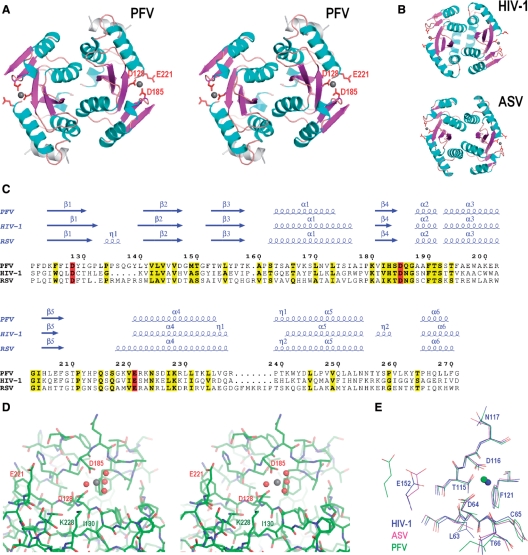
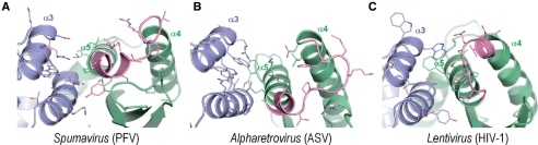
Establishment of the stable provirus is an essential step in retroviral replication, orchestrated by integrase (IN), a virus-derived enzyme. Until now, available structural information was limited to the INs of human immunodeficiency virus type 1 (HIV-1), avian sarcoma virus (ASV) and their close orthologs from the Lentivirus and Alpharetrovirus genera. Here, we characterized the in vitro activity of the prototype foamy virus (PFV) IN from the Spumavirus genus and determined the three-dimensional structure of its catalytic core domain (CCD). Recombinant PFV IN displayed robust and almost exclusively concerted integration activity in vitro utilizing donor DNA substrates as short as 16 bp, underscoring its significance as a model for detailed structural studies. Comparison of the HIV-1, ASV and PFV CCD structures highlighted both conserved as well as unique structural features such as organization of the active site and the putative host factor binding face. Despite possessing very limited sequence identity to its HIV counterpart, PFV IN was sensitive to HIV IN strand transfer inhibitors, suggesting that this class of inhibitors target the most conserved features of retroviral IN-DNA complexes.


Figures

Figure 1.
Figure 1.
Strand transfer activity of recombinant PFV IN. (A) Schematic of strand transfer reactions using circular DNA target. Concerted integration results in a gapped linear product, and the half-site process yields tailed relaxed circles. (B) Concerted integration activity of PFV INΔ in vitro. The enzyme (1.5 μM) was incubated with supercoiled pGEM target DNA in the absence (lane 1) or presence of 100 nM donor DNA (lanes 2–5). Blunt DNA fragments of varying lengths mimicking the U5 PFV cDNA terminus served as donors: 120-bp (lane 2), 420-bp (lane 3), an equimolar mixture of 120-bp and 420-bp donors (50 nM each, lane 4), or a 32-bp oligonucleotide (lane 5). Reaction products separated in 1.5% agarose gels were detected with ethidium bromide. Migration positions of concerted and half-site reaction products, open circular (o.c.) and supercoiled (s.c.) pGEM target DNA forms, donor DNA and a DNA size ladder are indicated. (C) PFV INΔ strand transfer activity in the presence of 0–2.5 μM blunt 32-bp donor DNA (lanes 2–10, as indicated). IN was omitted in lane 1. Here and on the remaining panels, the star indicates the migration position of the concerted integration product. (D) Time course of PFV INΔ strand transfer reactions in the presence of preprocessed (lanes 1–9) and blunt (lanes 10–18) 32-bp donor DNA. IN was omitted in lanes 1 and 10. (E) Comparison of strand transfer activities of full-length PFV IN and INΔ. Supercoiled pGEM target DNA was incubated with 0.38–3 μM INΔ (lanes 2–5) or full-length PFV IN (lanes 6–9) in the absence (lanes 2 and 6) or presence (lanes 3–5, 7–9) of preprocessed 32-bp donor substrate; IN was omitted in lane 1. (F) Effect of donor DNA length. Strand transfer reactions using PFV INΔ and preprocessed donors of 32–14 bp, as indicated atop the gel. The enzyme was omitted in lane 1 and donor DNA was omitted in lane 2.
Figure 2.
Figure 2.
Sequence analysis of concerted integration products. (A) Histograms of duplication size distributions in cloned concerted integration products from reactions mediated by PFV INΔ utilizing pre-processed 32-bp (black bars), 24-bp (light gray), or 16-bp (dark gray) donor DNA substrates. (B) A sequence logo representing nucleotide base frequencies at PFV integration sites. Target DNA sequences (n = 168) as joined to the reactive strand of donor DNA (from all 84 analyzed clones) were used in the alignment. Black arrowhead indicates the nucleotide joined to donor DNA (position 0 of the alignment); gray arrowhead indicates the insertion position of the second donor molecule into the complementary strand (position +3). The height of each logo is proportional to the frequency of the corresponding nucleotide within the alignment, while the height of each stack of logos reflects the level of conservation at each position. The figure was created using WebLogo (83).
Figure 3.
Figure 3.
PFV IN is sensitive to HIV IN strand transfer inhibitors. (A) Inhibition of the in vitro strand transfer activity by GS9137 and MK0518. Preprocessed 32-bp donor DNA substrate was incubated with PFV INΔ and supercoiled target DNA in the presence (lanes 1–5 and 7–11) or absence (lanes 6 and 12) of inhibitors. Final drug concentrations were 0.008 µM (lanes 1 and 7), 0.04 µM (lanes 2 and 8), 0.2 µM (lanes 3 and 9), 1 µM (lanes 4 and 10) and 5 µM (lanes 5 and 11). DMSO was present in all reactions at a final concentration of 1%. (B) Infection of D17 cells by PFV-based retroviral vector in the presence of strand transfer inhibitors MK0518 (black line and circles) or GS9137 (gray line and diamonds). Infectivity in the absence of inhibitors was set to 100%. The dotted gray line represents 50% inhibition.
Figure 4.
Figure 4.
Crystal structure of the PFV IN CCD. (A) Stereo view of the PFV CCD structure. Secondary structure elements are color-coded: light blue for α-helices, magenta for β-strands, and orange for loops; the C-terminal α-helix comprised of residues 293–304 is painted gray. The catalytic site residues Asp-128, Asp-185 and Glu-221 are shown as red sticks. Gray spheres are Mg atoms. (B) Structures of the HIV-1 and ASV IN CCDs (PDB accession codes 1bl3 and 1vsd, respectively) (C) Structure-based amino acid sequence alignment of PFV, HIV-1, and RSV IN CCDs. Secondary structure elements (α, alpha helix; β, beta strand; η, 310 helix) are shown above the alignment. Residue numbering corresponds to the full-length PFV IN. Residues conserved across the alignment are shown in bold and highlighted in yellow and those of the catalytic D,DX35E motif in red. (D) Stereo view of the PFV IN active site. Protein structure is shown as sticks; the Mg atom and the associated water molecules are gray and red spheres, respectively. Residues discussed in the text are labeled. (E) Superposition of the PFV, HIV-1 and ASV IN active sites. Carbon and Mg atoms (spheres) are colored according to the viral species: PFV, green; HIV-1, blue; ASV, magenta. Residue numbering corresponds to HIV-1 IN. (A), (B), (D) and (E) were created with PyMOL (DeLano Scientific, http://www.pymol.org), and (C) with ESPript (84).
Figure 5.
Figure 5.
Comparison of the putative co-factor binding faces of Spumavirus (PFV) (A) and Alpharetrovirus (ASV) (B) IN CCDs to the LEDGF-binding face of the Lentivirus (HIV-1) IN CCD (C). Dimeric CCD structures are shown as cartoons; protein chains are painted blue and green; helices α3, α4 and α5 are indicated, α4/5 connectors (40) are shown in magenta. Side chains of residues involved or potentially involved in co-factor binding are shown as sticks. The figure was created using PyMOL.

References

    1. Asante-Appiah E, Skalka AM. HIV-1 integrase: structural organization, conformational changes, and catalysis. Adv. Virus Res. 1999;52:351–369. - PubMed
    1. Craigie R. Retroviral DNA integration. In: Craig NL, Craigie R, Gellert M, Lambowitz AM, editors. Mobile DNA II. Washington DC: ASM Press; 2002. pp. 613–630.
    1. Lewinski MK, Bushman FD. Retroviral DNA integration—mechanism and consequences. Adv. Genet. 2005;55:147–181. - PubMed
    1. Sherman PA, Fyfe JA. Human immunodeficiency virus integration protein expressed in Escherichia coli possesses selective DNA cleaving activity. Proc. Natl Acad. Sci. USA. 1990;87:5119–5123. - PMC - PubMed
    1. Enssle J, Moebes A, Heinkelein M, Panhuysen M, Mauer B, Schweizer M, Neumann-Haefelin D, Rethwilm A. An active foamy virus integrase is required for virus replication. J. Gen. Virol. 1999;80:1445–1452. - PubMed

Publication types

MeSH terms

Associated data