. 2009 Jan;136(1):107-16.
doi: 10.1242/dev.026583. Epub 2008 Nov 26.

A SHH-responsive signaling center in the forebrain regulates craniofacial morphogenesis via the facial ectoderm

Affiliations

A SHH-responsive signaling center in the forebrain regulates craniofacial morphogenesis via the facial ectoderm

Diane Hu et al. Development. 2009 Jan.

Abstract

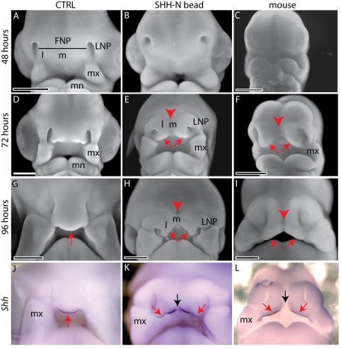
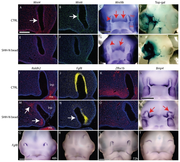
Interactions among the forebrain, neural crest and facial ectoderm regulate development of the upper jaw. To examine these interactions, we activated the Sonic hedgehog (SHH) pathway in the brain. Beginning 72 hours after activation of the SHH pathway, growth within the avian frontonasal process (FNP) was exaggerated in lateral regions and impaired in medial regions. This growth pattern is similar to that in mice and superimposed a mammalian-like morphology on the upper jaw. Jaw growth is controlled by signals from the frontonasal ectodermal zone (FEZ), and the divergent morphologies that characterize birds and mammals are accompanied by changes in the FEZ. In chicks there is a single FEZ spanning the FNP, but in mice both median nasal processes have a FEZ. In treated chicks, the FEZ was split into right and left domains that resembled the pattern present in mice. Additionally, we observed that, in the brain, fibroblast growth factor 8 (Fgf8) was downregulated, and signals in or near the nasal pit were altered. Raldh2 expression was expanded, whereas Fgf8, Wnt4, Wnt6 and Zfhx1b were downregulated. However, Wnt9b, and activation of the canonical WNT pathway, were unaltered in treated embryos. At later time points the upper beak was shortened owing to hypoplasia of the skeleton, and this phenotype was reproduced when we blocked the FGF pathway. Thus, the brain establishes multiple signaling centers within the developing upper jaw. Changes in organization of the brain that occur during evolution or as a result of disease can alter these centers and thereby generate morphological variation.


Figures

Fig. 1.
Fig. 1.
Activation of the SHH signaling pathway in the prosencephalon expands the ventral forebrain. Sections near the midline of each chick embryo were chosen for analysis based on the presence of or proximity to Rathke's pouch (RP). (A) In control embryos 24 hours after bead implantation (∼HH15), Nkx2.1 transcripts (green) were present in the diencephalon and telencephalon (arrows). Cells located in the optic recess also expressed Nkx2.1. (B) At this time, Shh (red) was expressed in the diencepahlon, but the expression domain did not extend beyond the optic recess (arrow). (C) In the brain, Pax6 (pink) was expressed in dorsal regions of the telencephalon, but was absent from the basal diencephalon. (D-F) In treated embryos, Nkx2.1 (D) and Shh (E) expression was expanded in the forebrain, while Pax6 (F) was repressed. Arrows indicate the dorsal location of the Pax6 expression domain. (G) Fgf8 (yellow) was expressed in the ectoderm (arrowhead) and forebrain (arrow) of controls at 24 hours after bead implantation. (H) At 60 hours, the ectoderm (arrowhead), brain (arrow) and optic recess (OR) expressed Fgf8. (I) By 72 hours, Fgf8 was expressed in the brain (arrow) and OR but was absent from the ectoderm. (J) At 24 hours after bead implantation, Fgf8 expression in the brain was mottled (arrow), and there was no expression in the optic recess. Expression of Fgf8 in the ectoderm appeared unaltered (arrowhead). (K) At 60 hours, Fgf8 was not expressed in the OR, and in the forebrain Fgf8 was downregulated. In the ectoderm of treated embryos, Fgf8 was not expressed (arrowhead). (L) By 72 hours, Fgf8 expression in the brain was maintained, but the OR domain was not apparent. Fgf8 was not expressed in the ectoderm at this time. Scale bar: 300 μm.
Fig. 2.
Fig. 2.
Altering the dorsoventral polarity of the brain perturbed facial development. (A) Control chick embryo 48 hours after bead implantation illustrating the medial (m) and lateral (l) regions of the FNP and their relationship with the lateral nasal processes (LNP). mn, mandibular process; mx, maxillary process. (B) In treated chick embryos, the nasal pits were not elongated, and the FNP was not well defined. (C) Growth of the upper jaw anlagen has not begun in mouse embryos at E9.5. (D) Control chick embryo 72 hours after bead implantation appeared normal. (E) Treated chick embryos had micro-opthalmia. In the FNP, growth occurred in lateral FNP (l; red arrows), whereas there was a lack of growth in the midline (m; arrowhead). (F) Growth of the middle and upper jaw in E11.0 mouse embryos occurred in lateral regions and began to form median nasal processes (arrows). Arrowhead indicates the median furrow present in the upper jaw of mouse embryos. (G) A control chick embryo 96 (∼HH28) hours post-implantation illustrates the normal morphology of the face. Arrow indicates growth in the medial portion of the FNP. (H,I) In treated chick embryos (H), growth of the FNP occurred in lateral domains (l; arrows) reminiscent of (I) median nasal processes (arrows) in mammals. Arrowheads indicate the median furrow in mouse embryos. (J) At HH28, Shh expression in the FEZ was restricted to a small domain located at the tip of the expanding FNP in chicks (red arrow, n=4). (K) In treated chicks, the Shh domain was divided into two halves that were larger than in normal chicks (red arrows; n=9/11). Black arrow indicates the lack of Shh expression in the midline. (L) In mice at E12 (n=6), Shh expression was evident in two large, distinct domains (red arrows), whereas in the midline, no Shh transcripts were observed (black arrow). The circled expression domain is in the basal forebrain. Scale bars: 1 mm in A,B,D,E,G,H,J,K; 500 μm in C,F,I,L.
Fig. 3.
Fig. 3.
The FEZ establishes proliferative zones in the mesenchyme of chick embryos. (A) Control embryo 72 hours after bead implantation. (B) Boxed area in B is shown in C. (C) BrDU incorporation illustrates the uniform distribution of proliferating cells across the FNP. (D) Treated embryos were malformed at 72 hours after bead implantation. (E) Boxed area is shown in F. (F) In treated embryos, fewer proliferating cells were detected in the midline. (G) The number of proliferating cells in midline (boxed areas) was reduced in treated embryos (n=10, P<0.05). Scale bar: 2 mm in A,D; 500 μm in B,E; 100 μm in C,F.
Fig. 4.
Fig. 4.
Morphology of the upper jaw skeleton. (A) Gross morphology of a control chick embryo at day 12 illustrates the relationship between the jaws. (B) In treated chick embryos, the upper jaw was shorter than in controls. Small eyes were also evident in these embryos. (C) A sagittal section through the distal tip of the upper jaw and stained with Milligan's modified Trichrome revealed the highly turbinated nasal capsule (arrow), the prenasal process of the premaxillary bone (arrowhead), the prenasal cartilage (pnc) and the egg tooth (asterisk). (D) In treated embryos, the nasal capsule (arrow) was rudimentary, but the egg tooth (*) was present. The prenasal process of the premaxillary bone was smaller and does not appear in this section. (E) Alcian Blue and Alizarin Red staining of a control head (day 13) illustrates the nasal bone (ns), jugal bar (Ju), maxillary bone (mx) and premaxillary bone (pmx). (F) In treated embryos, an ectopic cartilage nodule was apparent (arrow). The nasal bone, the maxillary bone and the jugal were greatly reduced in size, and the premaxillary bone appeared smaller. Scale bars: 2 mm in A,B; 100 μm in C,D; 1 mm in E,F.
Fig. 5.
Fig. 5.
Changes in signaling near the nasal pit in chick embryos. (A,B) Seventy-two hours after bead placement, Wnt4 (A, pink, arrow) and Wnt6 (B, green, arrow) were expressed in the epithelium of the nasal pit. (C) At this time, whole-mount in situ hybridization reveal Wnt9b expression throughout the epithelium covering the developing upper jaw (arrows), as well as in the lateral nasal (lnp) and maxillary (mxp) processes. However, Wnt9b was not expressed in the nasal epithelium (arrowhead). (D) X-gal reaction revealed activation of the canonical Wnt pathway throughout the developing FNP, lnp and mxp. (E,F) Wnt4 (E) and Wnt6 (F) were absent from the nasal epithelium in treated embryos 72 hours after implantation. (G) In treated embryos, Wnt9b transcripts spanned the mediolateral axis of the FNP (arrow). In these embryos, the nasal epithelium was exposed on the surface of the face and Wnt9b transcripts were absent from this tissue (arrowhead). (H) X-gal staining revealed that Wnt signaling still occurred in treated embryos. (I) Raldh2 was expressed (red) in the mesenchyme of the lnp and mxp 72 hours after bead implantation. (J) Fgf8 was expressed in the nasal epithelium of control embryos at this time. (K) Zfhx1b transcripts were present in the mesenchyme of the FNP in control embryos at this time. (L) Bmp4 transcripts were evident in the lateral regions of the FNP of control embryos 72 hours after bead implantation. (M) At this time Raldh2 was expressed (red) ectopically in mesenchyme of the FNP (arrows) and mxp. Expression in the lnp was not evident. (N) Fgf8 was absent form the medial nasal epithelium (arrow). (O) Zfhx1b was downregulated in the mesenchyme of treated embryos. (P) Bmp4 expression was expanded (red arrows) to surround the nasal pit in treated embryos at 72 hours. (Q) Fgf8 was expressed near the nasal pit in controls at 48 hours, but (R) in treated embryos Fgf8 was downregulated. (S) At 72 hours after bead placement, Fgf8 expression was strong surrounding the nasal pit, but (T) in treated embryos Fgf8 was downregulated. Scale bars: 250 μm in A,B,E,F,I-K,M-O; 1 mm in C,D,G,H,L,P,Q-T.
Fig. 6.
Fig. 6.
Blockade of FGF signaling may underlie skeletal changes after activation of SHH in the brain of chick embryos. (A) A control chick embryo illustrates morphology at day 13. (B) In treated embryos, the upper jaw was shorter than in controls and `clefts' were apparent (arrows). (C) Dorsal view of the head of a control stained with Alcian Blue and Alizarin Red at day 13 of development illustrates the nasal bone (nb), jugal bar (Ju), maxillary bone (mx) and premaxillary bone (pmx). The nasal capsule is indicated with an asterisk. The lower jaw is labeled mn. (D) The nasal bone (black nb), premaxillary bone (black pmx) and jugal bar (not shown) on the untreated side appeared normal. On the treated side, the premaxillary bone (red pmx) and nasal bone (red nb) appeared hypoplastic, and in this embryo the maxillary bone was absent. The nasal capsule (*) was greatly reduced on the treated side of embryos. The jugal bar is not in view. (E) Lateral view of control embryo illustrating the bones of the upper jaw. (F) Hypoplasia of the upper jaw skeleton was apparent in treated embryos. Scale bars: 2.5 mm in A,B; 2 mm in C-F.
Fig. 7.
Fig. 7.
Blockade of FGF signaling reduces cell proliferation in chick embryos. (A) Low magnification of the FNP after staining for BrdU. Boxes indicate locations of sections in B and C. (B,C) High magnification of cell proliferation data in regions near the bead (B) and on the contralateral side (C). (D) Graph illustrating proliferation in treated embryos (n=9, control 35.3%, s.d.=4.8%, treated 29.8%, s.d.=3.4%, P<0.05). Scale bars: 500 μm in A; 200 μm in B,C.

References

    1. Abzhanov, A. and Tabin, C. J. (2004). Shh and Fgf8 act synergistically to drive cartilage outgrowth during cranial development. Dev. Biol. 273, 134-148. - PubMed
    1. Abzhanov, A., Protas, M., Grant, B. R., Grant, P. R. and Tabin, C. J. (2004). Bmp4 and morphological variation of beaks in Darwin's finches. Science 305, 1462-1465. - PubMed
    1. Albrecht, U. E. G., Helms, J. A. and Lin, H. (1997). Visualization of gene expression patterns by in situ hybridization. In Molecular and Cellular Methods in Developmental Toxicology (ed. G. P. Daston), pp. 23-48. Boca Raton, FL: CRC Press.
    1. Ashique, A. M., Fu, K. and Richman, J. M. (2002). Endogenous bone morphogenetic proteins regulate outgrowth and epithelial survival during avian lip fusion. Development 129, 4647-4660. - PubMed
    1. Benouaiche, L., Gitton, Y., Vincent, C., Couly, G. and Levi, G. (2008). Sonic hedgehog signalling from foregut endoderm patterns the avian nasal capsule. Development 135, 2221-2225. - PubMed

Publication types

MeSH terms

Substances