

doi: 10.1242/dev.025908.
Epub 2008 Nov 26.
A new model for random X chromosome inactivation
Affiliations
- PMID: 19036804
- PMCID: PMC2630377
- DOI: 10.1242/dev.025908
Item in Clipboard
A new model for random X chromosome inactivation
Development.
2009 Jan.
Abstract
X chromosome inactivation (XCI) reduces the number of actively transcribed X chromosomes to one per diploid set of autosomes, allowing for dosage equality between the sexes. In eutherians, the inactive X chromosome in XX females is randomly selected. The mechanisms for determining both how many X chromosomes are present and which to inactivate are unknown. To understand these mechanisms, researchers have created X chromosome mutations and transgenes. Here, we introduce a new model of X chromosome inactivation that aims to account for the findings in recent studies, to promote a re-interpretation of existing data and to direct future experiments.
Figures
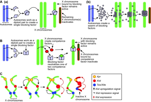
Three models of X chromosome inactivation. (A) The blocking
factor model. (a) Diploid autosomes work together to create a single
blocking factor (blue shape), which can bind to only one X chromosome,
preventing it from inactivating. (b) Nicodemi and Prisco used computer
simulations to show that if the autosomes produce a swarm of blocking factors
(blue dots) that can bind to each other and to the X chromosome, then all of
the blocking factor molecules will accumulate on a single X. (B) The
two factor model. Autosomes produce blocking factors and X chromosomes produce
transacting competence factors (red shape). Blocking factors bind to
competence factors with a two to one stoichiometry and then bind to one X
chromosome; the remaining competence factor binds to the other X, which
inactivates. (C) The sensing and choice model. After cells start to
differentiate, the two Xpr regions (yellow) pair and upregulate
Xist (red) on both X chromosmes. The Tsix/Xite region (blue)
pairs and chooses which chromosome to inactivate, and represses Xist
on the other. The X chromosome that represses Xist remains active
(green), and the other becomes inactive (red).

Stochastic model of X chromosome inactivation. (A) The
various components of the stochastic model. (B,a) Prior to cell
differentiation, the stochastic model proposes that the autosomes produce a
trans-acting factor (purple stars) that induces Tsix expression on
both X chromosomes (blue shape). (b) Tsix, when bound by
autosomal trans-acting factors, represses Xist transcription (red
shape). (c) After differentiation, an X-linked locus (yellow triangle)
produces a trans-acting factor (red circles) that attempts to upregulate
Xist. (d) Competition between Tsix- and
Xist-promoting factors creates a probability for each X chromosome to
inactivate. Cells that do not inactivate either X chromosome will continue to
produce the factor that promotes Xist upregulation in subsequent cell
cycles. Cells that inactivate both X chromosomes will either die, or
reactivate one. The percentages shown here reflect the proportions of each XCI
configuration observed 7 days after differentiation. Xa, active X chromosome;
Xi, inactive X chromosome.

Part of the mouse X chromosome inactivation center and sequence
replacements and deletions within it. Sequences used to replace portions
of the XIC are shown above the diagram, which shows their relative locations.
The effects of these replacements are listed in
Table 3. Below the diagram, the
dotted lines show XIC regions that have been deleted. The effects of these
deletions are listed in Table
2.

A problem with the two factor model. After XCI,
XTsixΔCpG:XTsixΔCpG
ES cells make three cell populations: one with a single active X (Xa)
chromosome and a single inactive X (Xi) chromosome; one with two Xa
chromosomes; and one with two Xi chromosomes. (A) Cells with normal XCI
result from blocking factor (BF) binding to one X chromosome and the
competence factor (C) binding to the other. (B) Cells with two Xa
chromosomes and two Xi chromosomes result from BF and C binding to the same X
chromosome. However, it is not clear how having both BF and C bound to a
single X chromosome results in these two different populations of cells.
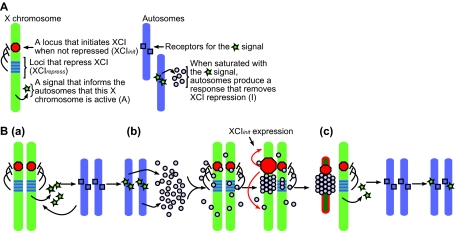
The feedback model of X chromosome inactivation. (A) A
description of the components required for the model. (B,a) The active
X chromosomes produce a trans-acting signal, A, that saturates specific sites
on the autosomes. (b) Once saturated, the autosomes produce a swarm of
inactivation signals, I. These signals bind to each other and to XCI
inhibitors on the X chromosomes. Once all of the XCI inhibitors on an X
chromosome are sufficiently bound by I, the XCI initiator induces
inactivation. (c) With only one active X chromosome producing A, the
autosomes are no longer saturated with A and stop producing I.
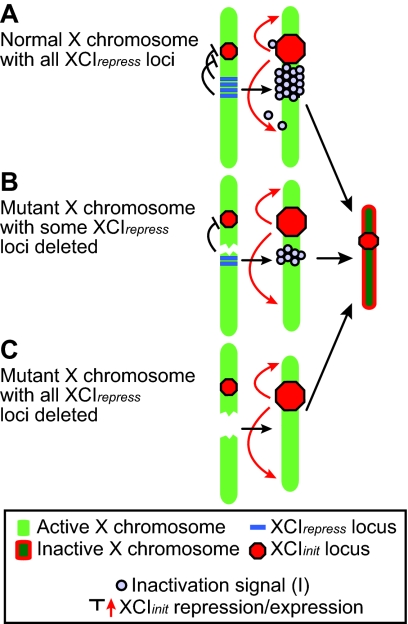
Three different paths to X inactivation. (A) In a normal X
chromosome, all of the XCIrepress loci must be turned off
by I. (B) In a mutant X chromosome where some of the
XCIrepress loci have been removed, only a small amount of
I is required to initiate XCI. Because these chromosomes have a lower
threshold to overcome before initiating XCI, there is a higher probability
that they will inactivate. (C) In a mutant X chromosome where all of
the XCIrepress loci have been removed, XCI will initiate
without any I.
References
-
- Augui, S., Filion, G. J., Huart, S., Nora, E., Guggiari, M., Maresca, M., Stewart, A. F. and Heard, E. (2007). Sensing X chromosome pairs before X inactivation via a novel X-pairing region of the Xic. Science 318, 1632-1636. - PubMed
-
- Bacher, C. P., Guggiari, M., Brors, B., Augui, S., Clerc, P., Avner, P., Eils, R. and Heard, E. (2006). Transient colocalization of X-inactivation centres accompanies the initiation of X inactivation. Nat. Cell Biol. 8, 293-299. - PubMed
-
- Borsani, G., Tonlorenzi, R., Simmler, M. C., Dandolo, L., Arnaud, D., Capra, V., Grompe, M., Pizzuti, A., Muzny, D., Lawrence, C. et al. (1991). Characterization of a murine gene expressed from the inactive X chromosome. Nature 351, 325-329. - PubMed
-
- Brockdorff, N., Ashworth, A., Kay, G. F., Cooper, P., Smith, S., McCabe, V. M., Norris, D. P., Penny, G. D., Patel, D. and Rastan, S. (1991). Conservation of position and exclusive expression of mouse Xist from the inactive X chromosome. Nature 351, 329-331. - PubMed
-
- Brown, C. J., Ballabio, A., Rupert, J. L., Lafreniere, R. G., Grompe, M., Tonlorenzi, R. and Willard, H. F. (1991a). A gene from the region of the human X inactivation centre is expressed exclusively from the inactive X chromosome. Nature 349, 38-44. - PubMed
Publication types
MeSH terms
Grants and funding
LinkOut - more resources
Full Text Sources
Other Literature Sources
