. 2009 Jan;174(1):91-100.
doi: 10.2353/ajpath.2009.080573. Epub 2008 Nov 26.

Type XVII collagen is a key player in tooth enamel formation

Affiliations

Type XVII collagen is a key player in tooth enamel formation

Takuya Asaka et al. Am J Pathol. 2009 Jan.

Abstract

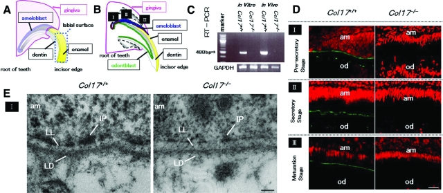
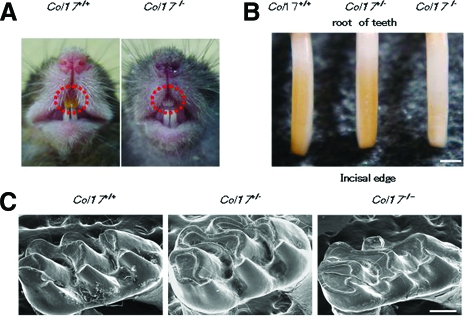
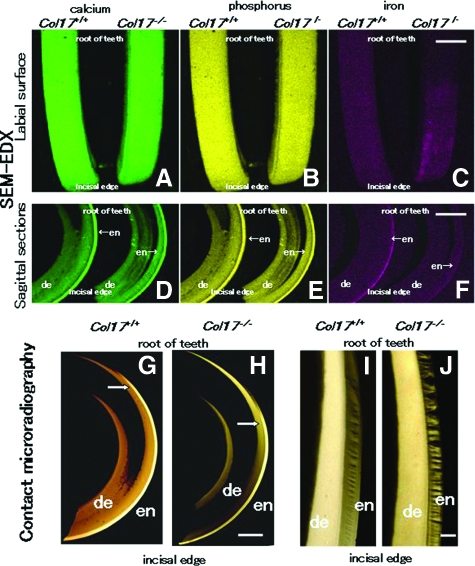
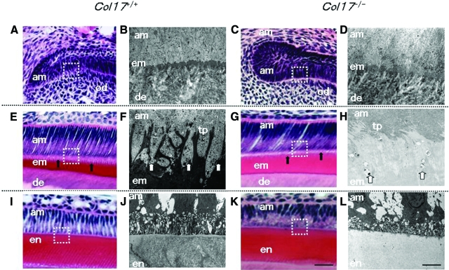
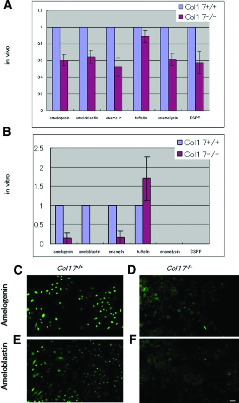
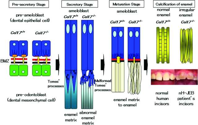
Inherited tooth enamel hypoplasia occurs due to mutations in genes that encode major enamel components. Enamel hypoplasia also has been reported in junctional epidermolysis bullosa, caused by mutations in the genes that encode type XVII collagen (COL17), a component of the epithelial-mesenchymal junction. To elucidate the pathological mechanisms of the enamel hypoplasia that arise from the deficiency of epithelial-mesenchymal junction molecules, such as COL17, we investigated tooth formation in our recently established Col17(-/-) and Col17 rescued mice. Compared with wild-type mice, the incisors of the Col17(-/-) mice exhibited reduced yellow pigmentation, diminished iron deposition, delayed calcification, and markedly irregular enamel prisms, indicating the presence of enamel hypoplasia. The molars of the Col17(-/-) mice demonstrated advanced occlusal wear. These abnormalities were corrected in the Col17 rescued humanized mice. Thus, the Col17(-/-) mice clearly reproduced the enamel hypoplasia in human patients with junctional epidermolysis bullosa. We were able to investigate tooth formation in the Col17(-/-) mice because the Col17(-/-) genotype is not lethal. Col17(-/-) mouse incisors had poorly differentiated ameloblasts that lacked enamel protein-secreting Tomes' processes and reduced mRNA expression of amelogenin, ameloblastin, and of other enamel genes. These findings indicated that COL17 regulates ameloblast differentiation and is essential for normal formation of Tomes' processes. In conclusion, COL17 deficiency disrupts the epithelial-mesenchymal interactions, leading to both defective ameloblast differentiation and enamel malformation.


Figures

Figure 1
Figure 1
COL17 expression in the tooth of Col17+/+ mice and COL17 absence in the tooth of Col17−/− mice. A, B: Mouse incisors are continuously elongating teeth. In the root of these incisors, ameloblasts (blue) and odontoblasts (green) secrete enamel matrix and dentin, respectively, during the secretory stage (II). I: the pre-secretory stage; II: the secretory stage; III: the maturation stage. C: A RT-PCR assay revealed that Col17 mRNA (488 bp band) was expressed in cultured ameloblasts from Col17+/+ mice (left lane) and Col17+/+ mouse teeth (second right). Col17 mRNA was not expressed in cultured ameloblasts from Col17−/− mice (second left lane) or Col17−/− mouse teeth (right hand lane). D: Immunofluorescence staining for COL17 (green) revealed that COL17 was expressed in the EMJ between ameloblasts and odontoblasts at the pre-secretory stage of a Col17+/+ mouse (upper, left), between ameloblasts and enamel matrix in the secretory stage (middle, left) and in the maturation stage (lower, left) of a Col17+/+ mouse. At the secretory stage, COL17 expression was weak, intermittent, or absent. In Col17−/− mice, no COL17 staining was observed in the EMJ at any stage (right column). am: ameloblast; od: odontblast. Scale bar = 20 μm. E: Ultrastructural features of the basement membrane zone at the pre-secretory stage. Normal hemidesmosomes were seen in the Col17+/+ mouse (left), but hypoplastic, malformed hemidesmosomes were observed in the Col17−/− mice (right). am: ameloblast; LL: lamina lucida; IP: inner attachment plaques; LD :lamina densa. Scale bar = 60 nm.
Figure 2
Figure 2
Dental phenotype of Col17−/− mice. A: At 4 weeks of age, a Col17−/− mouse (right) had whitish incisors. B: Incisors from Col17+/+ and Col17+/ mice showed yellowish color, although an incisor from a Col17−/− mouse seemed whitish (right). Scale bar = 500 μm. C: In the molars, tooth wear was more advanced for the Col17−/− mice (right) than for the Col17+/+ (left) and Col17+/ (center) mice. Scale bar = 250 μm.
Figure 3
Figure 3
Scanning electron microscopy of the sagittal section of maxillary incisors. A: A model of an upper incisor. The enamel layer indicated by an upper blue rectangle and by a lower red rectangle is enlarged in B, C, D and E, and in F, G, H and I, respectively. In the Col17−/− mouse, irregular inclinations of enamel rods without a normal network arrangement are observed (D, H), in contrast to the regular network of enamel rods observed in the Col17+/+ incisor (B, F) and in the Col17+/ incisor (C, G). The normal, regular network of enamel rods has been restored in the COL17 humanized mouse (E, I). en: enamel; de: dentin. Scale bar = 20 μm.
Figure 4
Figure 4
Difference in enamel formation between Col17+/+ and Col17−/− mice incisors. The labial surface (see Figure 1A) is featured in A, B, and C. A sagittal section is shown in D, E, and F. A, B: The labial surface of the maxillary incisors in both Col17+/+ (left) and Col17−/− (right) mice was scanned for calcium (green) and phosphorus (yellow) with EDX spectrometry. No obvious difference was observed in elemental distribution mapping. C: The same surfaces scanned in EDX for iron (red). In Col17−/− mice (right), the distribution of iron was irregular, compared with that of a Col17+/+ mice (left). Scale bar: (A, B, C) = 500 μm. D, E, F: The sagittal sections of the maxillary incisors of both Col17+/+ (left) and Col17−/− (right) mice scanned in EDX for calcium (green), phosphorus (yellow), and iron (red). No obvious difference is observed in the distribution of calcium or phosphorus between the Col17+/+ (left) and Col17−/− (right) mice. In the Col17−/− mice (right), the iron concentration in the enamel is lower than that in the Col17+/+ mouse (left; F). en: enamel; de: dentin. Scale bars in (D, E, F) = 1000 μm. G, H: Microradiographs of maxillary incisors in Col17+/+ (G) and Col17−/− (H) mice. The position (arrows) where sufficient mineralization occurred in the enamel judged from the low radio-opacity signal, moved toward the incisal edge in maxillary incisors of a Col17−/− mouse (G), compared with that in incisors of a Col17+/+ mouse (H). en: enamel; de: dentin. Scale bar: (G, H) = 500 μm. I, J: Microradiographs showing the mineralization pattern of the developing enamel at the maturation stage from Col17+/+ (I) and Col17−/− (J) mice. As compared with Col17+/+ (I), the mineralization demonstrated by radio-opacity of the enamel was irregular in both stages in Col17−/− mice (J), although there were no differences in the radio-opacity of dentine between Col17+/+ (I) and Col17−/− (J) mice. en: enamel; de: dentin. Scale bar: (I, J) = 100 μm.
Figure 5
Figure 5
Malformed Tomes’ processes and defective amelogenesis in Col17−/− mice A–D: At the pre-secretory and early secretory stages, the EMJ separates pre-ameloblasts and pre-odontoblasts. A, C: The overall structures of pre-ameloblasts and pre-odontoblasts were similar in the Col17+/+ (A) and Col17−/− (C) mice in the pre-secretory to the early secretory stages at the light microscopic level. B, D: Ultrastructurally, from the pre-secretory to the early secretory stages, the basement membrane between ameloblasts and odontoblasts was blurred in the Col17−/− mouse (D), compared with more obvious, intact basement membrane structures in a Col17+/+ mouse (B). E–H: At the secretory stage, Tomes’ processes are formed and enamel matrix is produced by ameloblasts. E, G: In the secretory stage, the processes of ameloblasts were malformed and blurred (arrows) in the Col17−/− mouse (G), compared with well-organized lattice-like structures of the Tomes’ processes (arrows) in the Col17+/+ mice (E). The thickness of the enamel matrix seemed similar both in Col17+/+ (E) and Col17−/− (G) mice. At the secretory stage, Tomes’ processes were apparently hypoplastic in the Col17−/− mouse (H), compared with normal Tomes’ processes in the Col17+/+ mouse (F). I–L: In the maturation stage, disruption of the processes of ameloblasts (am) was more advanced in the Col17−/− mouse (K), compared with regular processes in the Col17+/+ mice (I). At the maturation stage, the electron density of the enamel matrix is remarkably lower in the Col17−/− mouse (L) than that in the Col17+/+ mouse (J). In addition, enamel rod structures are blurred in the enamel matrix of the Col17−/− mouse (L). am: ameloblast; em: enamel matrix; en: enamel; de: dentin; od: odontoblast; tp: Tomes’ processes. Scale bars: (A, C, E, G, I, K) = 30 μm; (B, D, F, H, J, L) = 3 μm.
Figure 6
Figure 6
Expression of enamel proteins in Col17−/− ameloblasts. A: mRNA expression of all of the enamel proteins examined (amelogenin, ameloblastin, enamelin, tuftelin, enamelysin, and DSPP) was down-regulated in ameloblasts of incisors of the Col17−/−mice in vivo. B: In vitro ameloblasts cultured from incisors of the Col17−/− mice showed down-regulated mRNA expression of amelogenin, ameloblastin and enamelin, although tuftelin expression was up-regulated relative to tuftelin expression of the cultured ameloblasts from the Col17+/+ mice. Neither enamelysis nor DSPP was expressed in ameloblasts cultured from the Col17+/+ and Col17−/− mice. C: Protein expression (FITC, green) of amelogenin and ameloblastin was decreased in ameloblasts cultured from the Col17−/− mice (D, F), relative to that in ameloblasts cultured from the Col17+/+ mice (C, E). (C, D) amelogenin staining; (E, F) ameloblastin staining; (C, E) cells from Col17+/+ mice; (D, F) cells from Col17−/− mice. Scale bar = 20 μm.
Figure 7
Figure 7
Schemes of normal enamel formation in Col17+/+ mice and defective enamel formation in Col17−/− mice. In the Col17+/+ incisors (left), normal enamel matrix is formed by Tomes’ processes, resulting in intact enamel formation. In the Col17−/− incisors (right), disruptive Tomes’ processes produce disturbed enamel matrix, leading to irregular enamel formation.

References

    1. Maas R, Bei M. The genetic control of early tooth development. Crit Rev Oral Biol Med. 1997;8:4–39. - PubMed
    1. Liu F, Chu EY, Watt B, Zhang Y, Gallant NM, Andl T, Yang SH, Lu MM, Piccolo S, Schmidt-Ullrich R, Taketo MM, Morrisey EE, Atit R, Dlugosz AA, Millar SE. Wnt/beta-catenin signaling directs multiple stages of tooth morphogenesis. Dev Biol. 2008;313:210–224. - PMC - PubMed
    1. Borradori L, Sonnenberg A. Structure and function of hemidesmosomes: more than simple adhesion complexes. J Invest Dermatol. 1999;112:411–418. - PubMed
    1. McGrath JA, Gatalica B, Christiano AM, Li K, Owaribe K, McMillan JR, Eady RA, Uitto J. Mutations in the 180-kD bullous pemphigoid antigen (BPAG2), a hemidesmosomal transmembrane collagen (COL17A1), in generalized atrophic benign epidermolysis bullosa. Nat Genet. 1995;11:83–86. - PubMed
    1. Kirkham J, Robinson C, Strafford SM, Shore RC, Bonass WA, Brookes SJ, Wright JT. The chemical composition of tooth enamel in recessive dystrophic epidermolysis bullosa: significance with respect to dental caries. J Dent Res. 1996;75:1672–1678. - PubMed

Publication types

MeSH terms