

Beta-catenin is a mediator of the response of fibroblasts to irradiation
- PMID: 19036807
- PMCID: PMC2631337
- DOI: 10.2353/ajpath.2009.080576
Beta-catenin is a mediator of the response of fibroblasts to irradiation
Abstract
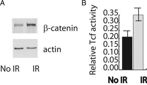
Radiation causes soft tissue complications that include fibrosis and deficient wound healing. beta-Catenin, a key component in the canonical Wnt-signaling pathway, is activated in fibrotic processes and wound repair and, as such, could play a role in mediating cellular responses to irradiation. beta-Catenin can form a transcriptionally active complex with members of the Tcf family. A reporter mouse model, in addition to human cell cultures, was used to demonstrate that ionizing radiation activates beta-catenin-mediated, Tcf-dependent transcription both in vitro and in vivo. Furthermore, radiation activates beta-catenin via a Wnt-mediated mechanism, as in the presence of dickkopf-1, an inhibitor of Wnt receptor activation, beta-catenin levels did not increase after irradiation. Fibroblast cell cultures were derived from mice expressing either null or stabilized beta-catenin alleles. Cells expressing stabilized beta-catenin alleles had a higher proliferation rate and formed more colony-forming units than wild-type or null cells after irradiation. Wound healing was studied in these same mice after irradiation. There was a positive correlation between the tensile strength of the wound, the expression levels of type 1 collagen in the skin, and beta-catenin levels. Mice treated with lithium showed increased beta-catenin levels and increased wound strength. beta-Catenin mediates the effects of ionizing radiation in fibroblasts, and its modulation has the potential to decrease the severity of radiation-induced soft tissue complications.
Figures


Similar articles
-
Beta-catenin signaling plays a disparate role in different phases of fracture repair: implications for therapy to improve bone healing.PLoS Med. 2007 Jul 31;4(7):e249. doi: 10.1371/journal.pmed.0040249. PLoS Med. 2007. PMID: 17676991 Free PMC article.
-
Bortezomib induces osteoblast differentiation via Wnt-independent activation of beta-catenin/TCF signaling.Blood. 2009 Apr 30;113(18):4319-30. doi: 10.1182/blood-2008-08-174300. Epub 2009 Feb 4. Blood. 2009. PMID: 19196662 Free PMC article.
-
Wnt/beta-catenin signaling in murine hepatic transit amplifying progenitor cells.Gastroenterology. 2007 Nov;133(5):1579-91. doi: 10.1053/j.gastro.2007.08.036. Epub 2007 Aug 28. Gastroenterology. 2007. PMID: 17983805
-
Interaction of nuclear receptors with the Wnt/beta-catenin/Tcf signaling axis: Wnt you like to know?Endocr Rev. 2005 Dec;26(7):898-915. doi: 10.1210/er.2003-0034. Epub 2005 Aug 26. Endocr Rev. 2005. PMID: 16126938 Review.
-
TCFs and Wnt/β-catenin signaling: more than one way to throw the switch.Curr Top Dev Biol. 2012;98:1-34. doi: 10.1016/B978-0-12-386499-4.00001-X. Curr Top Dev Biol. 2012. PMID: 22305157 Review.
Cited by
-
Regulation of early signaling and gene expression in the alpha-particle and bystander response of IMR-90 human fibroblasts.BMC Med Genomics. 2010 Jul 29;3:31. doi: 10.1186/1755-8794-3-31. BMC Med Genomics. 2010. PMID: 20670442 Free PMC article.
-
Proteomic analysis of radiation-induced changes in rat lung: Modulation by the superoxide dismutase mimetic MnTE-2-PyP(5+).Int J Radiat Oncol Biol Phys. 2010 Oct 1;78(2):547-54. doi: 10.1016/j.ijrobp.2010.03.037. Epub 2010 Jun 30. Int J Radiat Oncol Biol Phys. 2010. PMID: 20584581 Free PMC article.
-
Development of an easy-to-handle murine model for the characterization of radiation-induced gross and molecular changes in skin.Arch Plast Surg. 2018 Sep;45(5):403-410. doi: 10.5999/aps.2018.00101. Epub 2018 Sep 15. Arch Plast Surg. 2018. PMID: 30282410 Free PMC article.
-
Short- and long-term polystyrene nano- and microplastic exposure promotes oxidative stress and divergently affects skin cell architecture and Wnt/beta-catenin signaling.Part Fibre Toxicol. 2023 Jan 16;20(1):3. doi: 10.1186/s12989-023-00513-1. Part Fibre Toxicol. 2023. PMID: 36647127 Free PMC article.
-
Wnt signaling and injury repair.Cold Spring Harb Perspect Biol. 2012 Aug 1;4(8):a008078. doi: 10.1101/cshperspect.a008078. Cold Spring Harb Perspect Biol. 2012. PMID: 22723493 Free PMC article. Review.
References
-
- Nasonova E, Fussel K, Berger S, Gudowska-Nowak E, Ritter S. Cell cycle arrest and aberration yield in normal human fibroblasts. I. Effects of X-rays and 195 MeV u(-1) C ions. Int J Radiat Biol. 2004;80:621–634. - PubMed
-
- van Kampen M, Eble MJ, Lehnert T, Bernd L, Jensen K, Hensley F, Krempien R, Wannenmacher M. Correlation of intraoperatively irradiated volume and fibrosis in patients with soft-tissue sarcoma of the extremities. Int J Radiat Oncol Biol Phys. 2001;51:94–99. - PubMed
-
- Rodemann HP, Bamberg M. Cellular basis of radiation-induced fibrosis. Radiother Oncol. 1995;35:83–90. - PubMed
-
- O'Sullivan B, Levin W. Late radiation-related fibrosis: pathogenesis, manifestations, and current management. Semin Radiat Oncol. 2003;13:274–289. - PubMed
-
- Gavert N, Ben-Ze'ev A. Beta-catenin signaling in biological control and cancer. J Cell Biochem. 2007;102:820–828. - PubMed
Publication types
MeSH terms
Substances
LinkOut - more resources
Full Text Sources
Other Literature Sources
Molecular Biology Databases
