. 2009 Feb;83(4):1992-2003.
doi: 10.1128/JVI.01621-08. Epub 2008 Nov 26.

Distinct domains within APOBEC3G and APOBEC3F interact with separate regions of human immunodeficiency virus type 1 Vif

Affiliations

Distinct domains within APOBEC3G and APOBEC3F interact with separate regions of human immunodeficiency virus type 1 Vif

Rebecca A Russell et al. J Virol. 2009 Feb.

Abstract

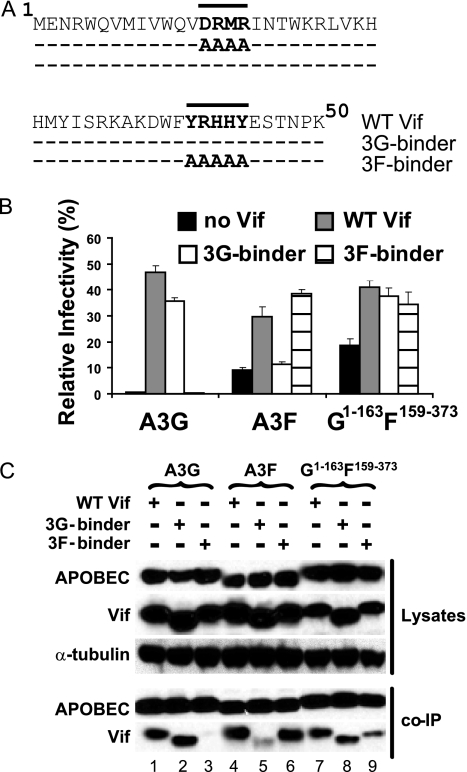
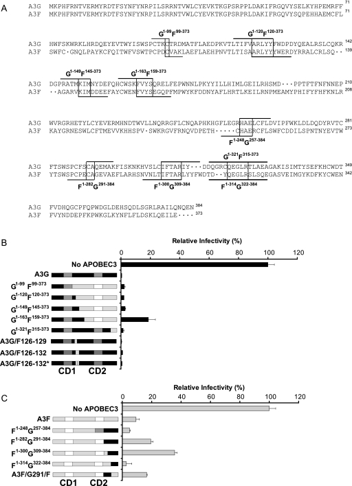
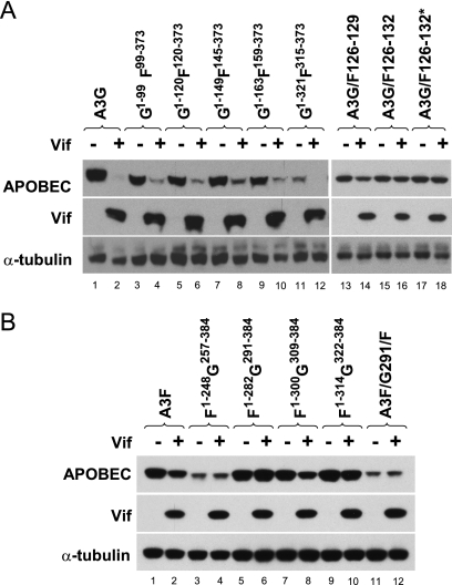
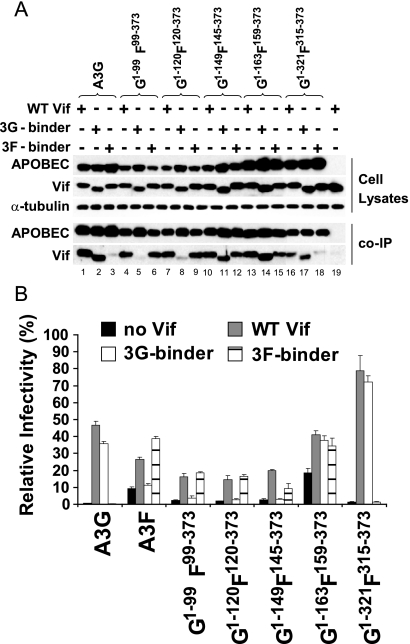
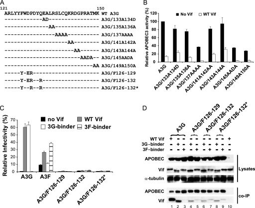
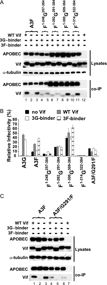
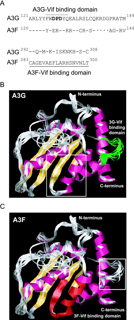
Human APOBEC3G (A3G) and APOBEC3F (A3F) inhibit the replication of Vif-deficient human immunodeficiency virus type 1 (HIV-1). HIV-1 Vif overcomes these host restriction factors by binding to them and inducing their degradation. Thus, the Vif-A3G and Vif-A3F interactions are attractive targets for antiviral drug development, as inhibiting these interactions could allow the host defense mechanism to control HIV-1 replication. Recently, it has been reported that amino acids 105 to 156 of A3G are involved in the interaction with Vif; however, to date, the region of A3F involved in Vif binding has not been identified. Using our previously reported Vif mutants that are capable of binding to only A3G (3G binder) or only A3F (3F binder), in conjunction with a series of A3G-A3F chimeras, we have now mapped the APOBEC3-Vif interaction domains. We found that the A3G domain that interacts with the Vif YRHHY region is located between amino acids 126 and 132 of A3G, which is consistent with the conclusions reported in previous studies. The A3F domain that interacts with the Vif DRMR region did not occur in the homologous domain but instead was located between amino acids 283 and 300 of A3F. These studies are the first to identify the A3F domain that interacts with the Vif DRMR region and show that distinct domains of A3G and A3F interact with different Vif regions. Pharmacological inhibition of either or both of these Vif-A3 interactions should prevent the degradation of the APOBEC3 proteins and could be used as a therapy against HIV-1.


Figures

FIG. 1.
FIG. 1.
The Vif interaction domain is located in the N terminus of A3G and the C terminus of A3F. (A) Schematic representation of Vif mutants defective in A3G and A3F binding. The top line depicts the first 50 amino acids of WT Vif, the second line shows the four alanine substitutions made in the 3G binder (DRMR>A4), and the third line shows the five alanine substitutions made in the 3F binder (YRHHY>A5). Dashes indicate identical amino acids. (B) Effect of A3G, A3F, and the G1-163F159-373 chimera on HIV-1 infectivity in the presence and absence of Vif. To determine APOBEC3 sensitivity to WT and mutant Vif proteins, 293T cells were transfected with the HIV-1 vector pHDV-EGFP, a vesicular stomatitis virus glycoprotein expression plasmid (pHCMV-G), an APOBEC-expressing plasmid, and a Vif-expressing plasmid (WT Vif, the 3G binder, or the 3F binder). HIV-1-derived vector-enhanced green fluorescent protein (EGFP) virus was also prepared in the presence of the APOBEC3 proteins but in the absence of any Vif proteins to determine the function of the APOBEC3 proteins. As a positive control, HDV-EGFP virus was prepared in the absence of any APOBEC3 proteins. The infectivity of the virus produced from the transfected cells harvested after 48 h was determined by infection of TZM-bl indicator cells and quantitation of the resulting luciferase enzyme activity. The data shown are plotted as the infectivity relative to that produced in the absence of any APOBEC3 proteins (not shown), which was set to 100%, with standard errors of the means (SEM) from three independent experiments. (C) Co-IP assays to determine binding of Vif and APOBEC3 proteins. To assess the level of binding between the APOBEC3 proteins and the WT and mutant Vif proteins, 293T cells were cotransfected with N-terminally FLAG-tagged versions of A3G, A3F, or G1-163F159-373 along with WT Vif, the 3G binder, or the 3F binder. The transfected cell lysates were analyzed for the expression of the APOBEC3 and Vif proteins using anti-FLAG and anti-Vif antibodies, respectively, by Western blotting. The cell lysates were also analyzed for α-tubulin to control for the amount of cell lysate examined. Additionally, the cell lysates were analyzed in an immunoprecipitation assay using anti-FLAG resin to immunoprecipitate the FLAG-tagged APOBEC3 proteins. The immunoprecipitated proteins were then analyzed by Western blotting using anti-FLAG and anti-Vif antibodies. A representative analysis is shown.
FIG. 2.
FIG. 2.
Inhibition of HIV-1 ΔVif by the A3G/A3F chimeras. (A) Schematic representation of the crossover points of each chimera. Alignments of the A3G and A3F amino acid sequences and the crossover points in each chimera are shown. Identical amino acids in A3G and A3F at the crossover junctions are shown in boxes; dots indicate gaps in the sequence. (B) Effects of the GF and A3G/F126-129, A3G/F126-132, and A3G/F126-132* chimeras on HIV-1 infectivity. A3G is depicted in black, and A3G CD1 and CD2 are depicted in dark gray. A3F is depicted in light gray, and the A3F CD1 and CD2 are depicted in white. To determine GF chimera function, 293T cells were transfected with pHDV-EGFP, pHCMV-G, and an APOBEC-expressing plasmid. As a positive control, HDV-EGFP virus was prepared in the absence of any APOBEC3 proteins. After 48 h, the infectivity of the virus produced from the transfected cells was determined by infection of TZM-bl indicator cells and quantitation of the resulting luciferase enzyme activity. The data shown are plotted as the infectivity relative to that produced in the absence of any APOBEC3 proteins, which was set to 100%, with SEM from three independent experiments. (C) Effects of the FG and A3F/G291/F chimeras on HIV-1 infectivity. The color scheme and experimental protocol is the same as that described above.
FIG. 3.
FIG. 3.
Expression levels and Vif sensitivity of A3G-A3F chimeras. (A) Expression of A3G-A3F chimeras in the absence or presence of Vif. The GF chimera-expressing plasmids and WT Vif expression plasmids were cotransfected into 293T cells. An APOBEC3-to-Vif expression plasmid molar ratio of 1:5 was used for the cotransfections. The transfected cell lysates were analyzed by Western blotting for the expression of the APOBEC3 and Vif proteins using anti-FLAG and anti-Vif antibodies, respectively. The cell lysates were also analyzed for tubulin expression using an anti-tubulin antibody to control for the amount of cell lysate examined. (B) Expression of A3F-A3G chimeras in the presence and absence of Vif. The FG chimera-expressing plasmids and WT Vif-expressing plasmid were cotransfected into 293T cells. An APOBEC3-to-Vif molar ratio of 1:10 was used for the cotransfections. The APOBEC3, Vif, and tubulin proteins were detected by Western blotting as described in the text.
FIG. 4.
FIG. 4.
The A3G Vif-binding domain is localized to amino acids 121 to 149. (A) Co-IP assays to determine binding of Vif and GF chimeras. Binding of the GF chimeras to WT Vif, the 3G binder, and the 3F binder was determined as described in the legend of Fig. 1C by co-IP and Western blotting. A representative analysis is shown. (B) Effects of GF chimeras on HIV-1 infectivity and their sensitivity to Vif proteins. HIV-1 infections of TZM-bl cells were carried out as described in the legend of Fig. 1B. The SEM determined from two to four independent experiments are shown.
FIG. 5.
FIG. 5.
Amino acids 126 to 132 of A3G are critical for binding to Vif. (A) Schematic representation of mutants of the Vif-binding domain of A3G. The top line depicts amino acids 121 to 150 of WT A3G, the next seven lines depict the alanine and aspartic acid substitution mutants, and the last three lines depict the A3G/F126-129, A3G/F126-132, and A3G/F126-132* chimeras; dashes indicate identical amino acids. (B) Effects of alanine substitution mutations on cytidine deaminase activity. The alanine substitution mutants shown in A were transfected into 293T cells in the presence or absence of Vif, the cell lysates were harvested 48 h posttransfection, and cytidine deaminase activity was determined using 0.2 μg of total protein. The data shown are plotted as the level of A3G activity relative to that obtained from WT A3G in the absence of any Vif, which was set to 100%. The SEM determined for two to three independent experiments are shown. (C) Effects of A3G/F chimeras on HIV-1 infectivity in the presence or absence of Vif. The chimeras A3G/F126-129, A3G/F126-132, and A3G/F126-132* were tested for their sensitivity to WT Vif and both the 3G and 3F binders as described in the legend of Fig. 1B. The SEM determined for two to four independent experiments are shown. (D) Co-IP assays to determine binding of Vif and A3G/F chimeras. The binding of the A3G/F126-129, A3G/F126-132, and A3G/F126-132* chimeras to the 3G binder and the 3F binder was determined as described in the legend of Fig. 1C by co-IP and Western blotting. A representative analysis is shown.
FIG. 6.
FIG. 6.
Amino acids 283 to 300 of A3F are critical for binding to Vif. (A) Co-IP assays to determine binding of Vif to the FG chimeras. The binding of the FG chimeras to the 3G binder and the 3F binder was determined as described in the legend of Fig. 1C by co-IP and Western blotting. A representative analysis is shown. (B) Effects of the FG and A3F/G291/F chimeras on HIV-1 infectivity and their sensitivity to Vif proteins. Determination of the Vif sensitivity of the FG and A3F/G291/F chimeras was carried out as described in the legend of Fig. 1B. The SEM determined for two to four independent experiments are shown. (C) Co-IP assays to determine binding of Vif to the A3F/G291/F chimera. The binding of the A3F/G291/F chimera to WT Vif, the 3G binder, and the 3F binder was determined as described in the legend of Fig. 1C by co-IP and Western blotting. A representative analysis is shown.
FIG. 7.
FIG. 7.
Sequence and model structure of Vif binding domains of A3G and A3F. (A) Alignment of A3G and A3F sequences within the A3G Vif- and A3F Vif-binding sites. Sequence alignment of the A3G Vif-binding domain with the corresponding region in A3F and the A3F Vif-binding domain with the corresponding region in A3G. Dashes indicate identical amino acids, and dots indicate gaps in the sequence. The residues in boldface type are those previously shown to be critical for Vif binding. (B) Structure of the C-terminal domain of A3G and location of the N-terminal Vif-binding domain mapped onto the corresponding region (shown in green). The highlighted residues span amino acids 315 to 322 of A3G, which correspond to amino acids 126 to 132 of A3G. The location in the C terminus of the A3F Vif-binding domain is shown within the boxed area. β-Sheets 1 to 5 are shown in pink, and α-helices 1 to 5 are shown in yellow. (C) Structure of the C-terminal domain of A3G and location of the Vif-binding domain of A3F mapped onto the corresponding region (shown in green). The highlighted residues span amino acids 292 to 308 of the C terminus of A3G, which correspond to amino acids 283 to 300 of A3F. The location of the N-terminal A3G Vif-binding domain is shown within the boxed area. β-Sheets 1 to 5 are shown in pink, and α-helices 1 to 5 are shown in yellow.

Similar articles

Cited by

References

    1. Bishop, K. N., R. K. Holmes, and M. H. Malim. 2006. Antiviral potency of APOBEC proteins does not correlate with cytidine deamination. J. Virol. 808450-8458. - PMC - PubMed
    1. Bishop, K. N., R. K. Holmes, A. M. Sheehy, N. O. Davidson, S. J. Cho, and M. H. Malim. 2004. Cytidine deamination of retroviral DNA by diverse APOBEC proteins. Curr. Biol. 141392-1396. - PubMed
    1. Bogerd, H. P., B. P. Doehle, H. L. Wiegand, and B. R. Cullen. 2004. A single amino acid difference in the host APOBEC3G protein controls the primate species specificity of HIV type 1 virion infectivity factor. Proc. Natl. Acad. Sci. USA 1013770-3774. - PMC - PubMed
    1. Boussif, O., F. Lezoualc'h, M. A. Zanta, M. D. Mergny, D. Scherman, B. Demeneix, and J. P. Behr. 1995. A versatile vector for gene and oligonucleotide transfer into cells in culture and in vivo: polyethylenimine. Proc. Natl. Acad. Sci. USA 927297-7301. - PMC - PubMed
    1. Chen, K. M., E. Harjes, P. J. Gross, A. Fahmy, Y. Lu, K. Shindo, R. S. Harris, and H. Matsuo. 2008. Structure of the DNA deaminase domain of the HIV-1 restriction factor APOBEC3G. Nature 452116-119. - PubMed

Publication types

MeSH terms

Substances

LinkOut - more resources