. 2008 Nov 26;28(48):12815-9.
doi: 10.1523/JNEUROSCI.2665-08.2008.

Disruption of Nectin-like 1 cell adhesion molecule leads to delayed axonal myelination in the CNS

Affiliations

Disruption of Nectin-like 1 cell adhesion molecule leads to delayed axonal myelination in the CNS

Jinsil Park et al. J Neurosci. .

Abstract

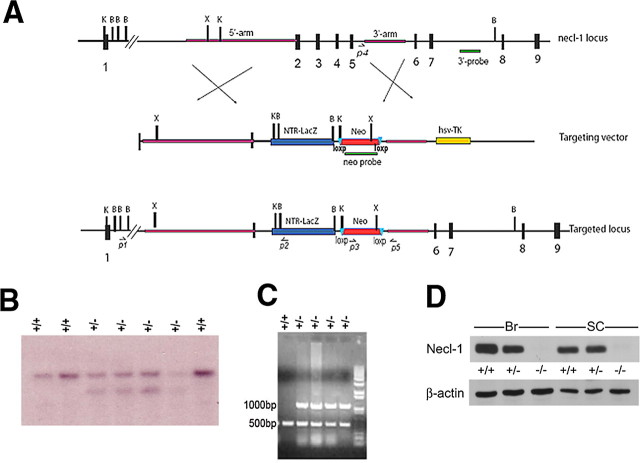
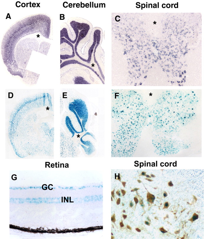
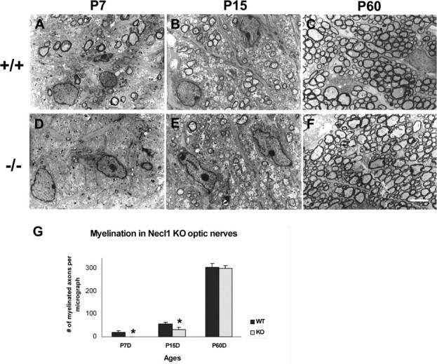
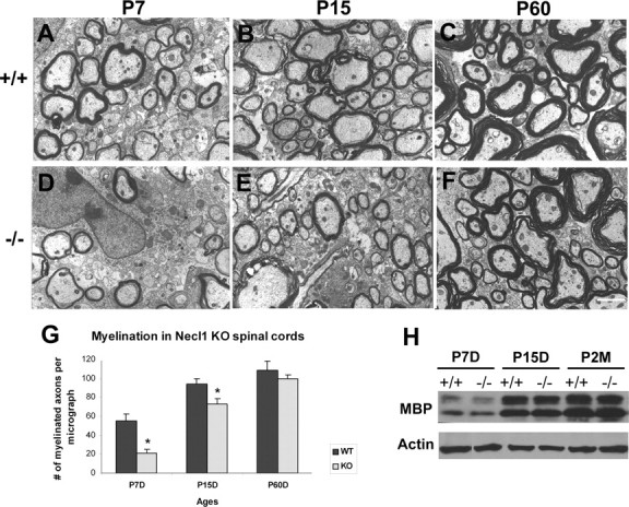
Nectin-like 1 (Necl-1) is a neural-specific cell adhesion molecule that is expressed in both the CNS and PNS. Previous in vitro studies suggested that Necl-1 expression is essential for the axon-glial interaction and myelin sheath formation in the PNS. To investigate the in vivo role of Necl-1 in axonal myelination of the developing nervous system, we generated the Necl-1 mutant mice by replacing axons 2-5 with the LacZ reporter gene. Expression studies revealed that Necl-1 is exclusively expressed by neurons in the CNS. Disruption of Necl-1 resulted in developmental delay of axonal myelination in the optic nerve and spinal cord, suggesting that Necl-1 plays an important role in the initial axon-oligodendrocyte recognition and adhesion in CNS myelination.


Figures

Figure 1.
Figure 1.
Gene targeting of Necl-1. A, The wild-type Necl-1 genomic organization, targeting vector, and the targeted allele after the predicted homologous recombination event are shown. The 3′ probe used for Southern blot analysis is shown below the wild-type genomic map. Primers (P3, P4 and P5) for genotyping are indicated. B, BamH1; C, ClaI; E, EcoRI; X, XhoI. B, Genotyping of F1 animals by Southern blot analysis with the 3′ probe. The wild-type allele is 10 kb and the mutant allele 8.0 kb. The genotype of each sample is indicated on top of the gel. C, Genotyping of F1 animals by PCR with three primers P3, P4 and P5 as indicated in A. D, Western immunoblotting of adult brain (Br) and spinal cord (SC) tissues with anti-Necl-1 polyclonal antibody. Genotypes of animals were indicated at the top of the lanes.
Figure 2.
Figure 2.
Restrictive expression of Necl-1 in adult CNS neurons. A–F, Cerebral, spinal and cerebellar tissues were isolated from Necl-1 heterozygous animals and subjected to in situ RNA hybridization with Necl-1 probe (A–C) or LacZ staining (D–F). Asterisks indicate the lack of Necl-1 expression in the white matter of the cerebral cortex (A, D), cerebellum (B, E) and spinal cord (C, F). G, LacZ staining in retinal ganglionic neurons (GC) and inner nuclear layer (INL) in the adult heterozygous mice. H, Double labeling of LacZ signal and anti-NeuN immunohistochemical staining.
Figure 3.
Figure 3.
Delayed axonal myelination in the Necl-1 mutant optic nerves. A–F, Ultra-thin sections from P7, P15 and P60 wild-type (A–C) and Necl-1 mutant (D–F) optical nerves were examined under transmission EM. G, Number of myelinated axons per micrograph (500 μm2) in the wild-type and mutant tissues at various postnatal stages (n = 3). Statistically significant differences (p value <0.05) between the wild-type and mutant animals were indicated by asterisks. Scale bar, 2 μm.
Figure 4.
Figure 4.
Delayed axonal myelination in the Necl-1 mutant spinal cords. A–F, Lateral white matter tissues from P7, P15 and P60 wild-type (A–C) and Necl-1 mutant (D–F) spinal cords were processed for EM analyses. G, Number of myelinated axons per micrograph (500 μm2) in the wild-type and mutant tissues at various postnatal stages (n = 3). Statistically significant differences (p <0.05) between the wild-type and mutant animals were indicated by asterisks. Scale bar, 2 μm. H, Western immunoblotting of P7–P60 spinal cords with anti-MBP and anti-β-actin antibodies.

Similar articles

Cited by

References

    1. Biederer T. Bioinformatic characterization of the SynCAM family of immunoglobulin-like domain-containing adhesion molecules. Genomics. 2006;87:139–150. - PubMed
    1. Biederer T, Sara Y, Mozhayeva M, Atasoy D, Liu X, Kavalali ET, Südhof TC. SynCAM, a synaptic adhesion molecule that drives synapse assembly. Science. 2002;297:1525–1531. - PubMed
    1. Colello RJ, Pott U. Signals that initiate myelination in the developing mammalian nervous system. Mol Neurobiol. 1997;15:83–100. - PubMed
    1. Colognato H, Ramachandrappa S, Olsen IM, ffrench-Constant C. Integrins direct Src family kinases to regulate distinct phases of oligodendrocyte development. J Cell Biol. 2004;167:365–375. - PMC - PubMed
    1. Edgar JM, Garbern J. The myelinated axon is dependent on the myelinating cell for support and maintenance: molecules involved. J Neurosci Res. 2004;76:593–598. - PubMed

Publication types

MeSH terms

LinkOut - more resources