. 2008 Nov 26;28(48):12901-12.
doi: 10.1523/JNEUROSCI.4629-08.2008.

Traumatic brain injury-induced hippocampal neurogenesis requires activation of early nestin-expressing progenitors

Affiliations

Traumatic brain injury-induced hippocampal neurogenesis requires activation of early nestin-expressing progenitors

Tzong-Shiue Yu et al. J Neurosci. .

Abstract

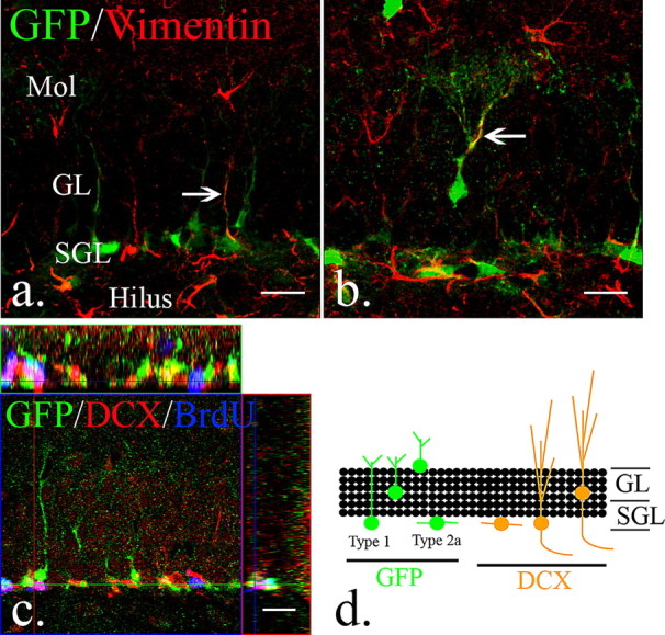
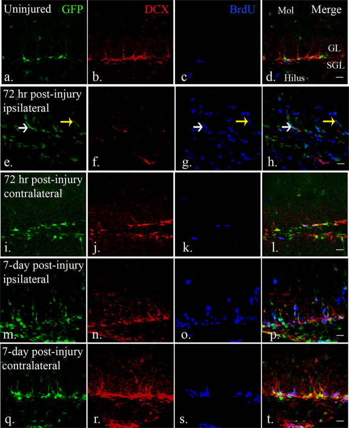
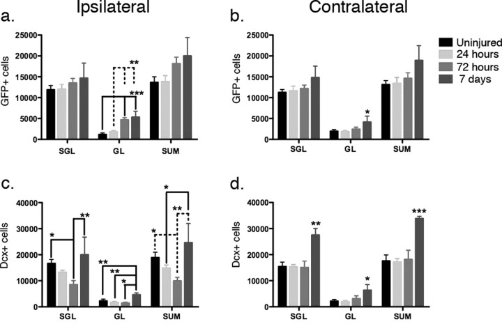
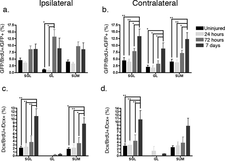
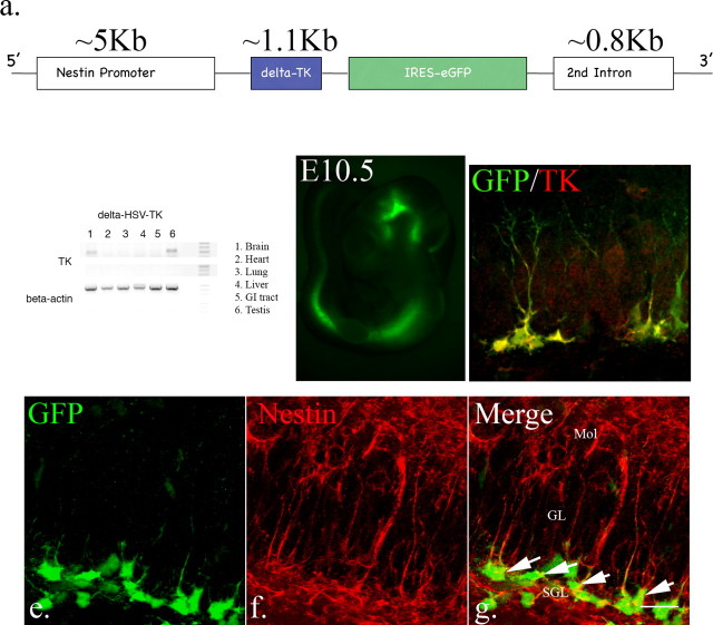
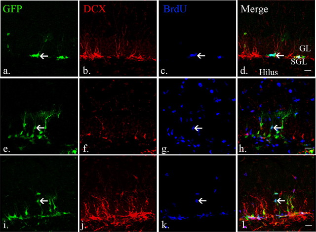
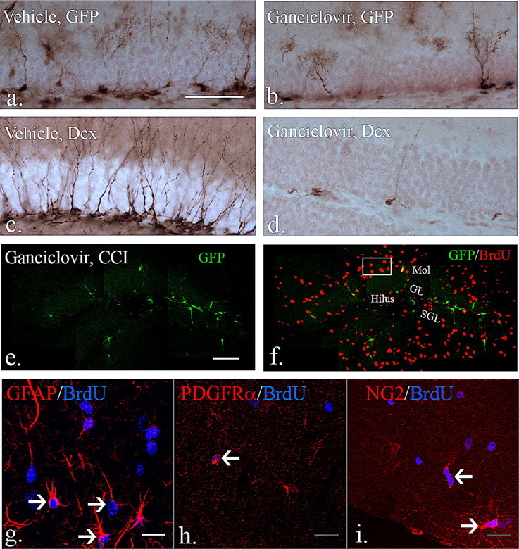
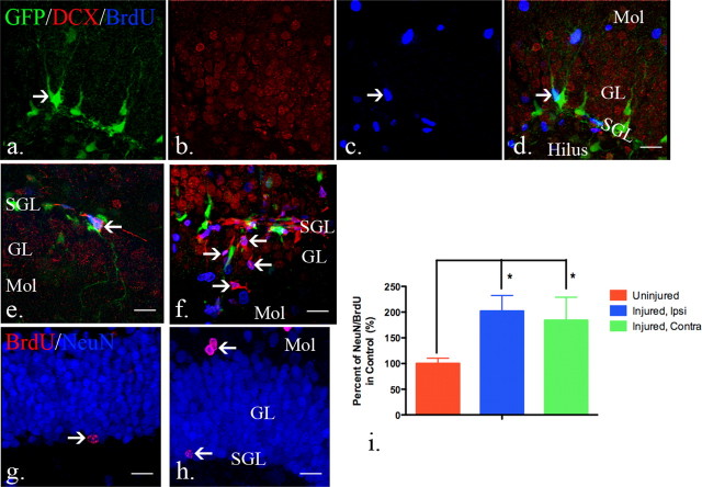
It is becoming increasingly clear that brain injuries from a variety of causes stimulate neurogenesis within the hippocampus. It remains unclear, however, how robust this response may be and what primary cell types are involved. Here, using a controlled cortical impact model of traumatic brain injury on a previously characterized transgenic mouse line that expresses enhanced green fluorescent protein (eGFP) under the control of the nestin promoter, we demonstrate that it is the earliest type-1 quiescent progenitor cells that are induced to proliferate and migrate outside the subgranular layer of the dentate gyrus. This type-1 cell activation occurs at the same time that we observe adjacent but more differentiated doublecortin-expressing progenitors (type-2 cells) being eliminated. Also, although type-2 cells remain intact in the contralateral (uninjured) dentate gyrus, the type-1 cells there are also activated and result in increased numbers of the doublecortin-expressing type-2 cells. In addition, we have generated a novel mouse transgenic that expresses a modified version of the herpes simplex virus thymidine kinase along with eGFP that allows for the visualization and inducible ablation of early dividing progenitors by exposing them to ganciclovir. Using this transgenic in the context of traumatic brain injury, we demonstrate that these early progenitors are required for injury-induced remodeling to occur. This work suggests that injury-induced hippocampal remodeling following brain injury likely requires sustained activation of quiescent early progenitors.


Figures

Figure 1.
Figure 1.
The distribution and types of neural progenitors in the adult dentate gyrus. a, A subset of eGFP-expressing cells in the subgranular layers express putative neural stem cell markers, like vimentin (arrow). b, However, vimentin and eGFP-expressing cells were also occasionally observed in the granular layers (arrow). c, In the adult dentate gyrus, early eGFP-expressing cells are distinguishable from BrdU/DCX-positive fast-dividing type 2b neural progenitors. d, The scheme represents the distribution of eGFP-expressing type 1 and 2a neural progenitors and DCX-expressing type 2b and 3 neural progenitors in the dentate gyrus of the hippocampus. Scale bars, 20 μm. Mol, Molecular layer; SGL, subgranular layer; GL, granular layer.
Figure 2.
Figure 2.
The distribution and response of neural progenitors in the adult dentate gyrus after CCI injury. a–d, eGFP- and DCX-expressing cells were found in the subgranular and granular layers of the dentate gyrus. BrdU-labeled cells were rarely seen in the dentate gyrus in uninjured mice. (e–h, After injury (72 h), eGFP-expressing early neural progenitors were detected in the subgranular layers (white arrow), as well as granular layers (yellow arrow). There is an apparent decrease of DCX-expressing late neural progenitors in the dentate gyrus in the ipsilateral hemispheres (f) at the same time that abundant BrdU-labeled cells were found in the dentate gyrus (g). (i–l) In the contralateral dentate gyrus, there is minimal apparent activation of GFP-expressing cells that mimics the control dentate gyrus. m–p, Robust numbers of eGFP-expressing cells persist in the ipsilateral dentate gyrus 7 d after injury (m). DCX-expressing late neural progenitors are more recovered at this time point (n) and there remains a large number of BrdU-positive cells (o). q–t, There is also an apparent increase in cell number and proliferation of both eGFP (q) and DCX-expressing (r) cells in the contralateral dentate gyrus 7 d after injury. Scale bars, 20 μm. Mol, Molecular layer; SGL, subgranular layer; GL, granular layer.
Figure 3.
Figure 3.
Quantification of early and late neural progenitors in the dentate gyrus after injury. In the uninjured brains, there were similar numbers of eGFP-expressing cells in the dentate gyrus in both hemispheres (16,647 ± 3046 and 15,417 ± 3407 in the subgranular layers, 2259 ± 1370 and 2169 ± 1266 in the granular layers; mean ± SD). a, In the ipsilateral dentate gyrus, no significant change was observed in the early neural progenitors in the subgranular layers (SGL). A significant increase in the number of early neural progenitors was observed in the granular layers (GL) (p = 0.0001, one-way ANOVA). Post hoc analysis revealed a significant increase of early neural progenitors 72 h and 7 d after CCI injury (p < 0.01) when compared with uninjured samples and 24 h after injury and the increase in the granular layers contributed to a significant increase in total neural progenitor number (p = 0.0474, one-way ANOVA). b, A similar phenomenon was noted in the contralateral dentate gyrus, with no significant change in the subgranular layers but increases in the granular layers (p = 0.1221, 0.0229, and 0.0433 in subgranular layers, granular layers and total (SUM), respectively, one-way ANOVA). c, The opposite result was seen in the DCX-expressing late neural progenitor population where in the ipsilateral dentate gyrus, there was a significant difference in both layers examined (p = 0.0116, 0.0016 and 0.0050 for DCX-expressing cell number in subgranular layers, granular layers and total). In the subgranular layer, there was a significant decrease in late neural progenitors 72 h after the injury compared with 7 d after the injury, when the cell population had returned to baseline (p < 0.05 with post hoc Newman–Keuls multiple comparison test). The granular layer revealed a similar increase 7 d after the injury when compared with uninjured group, 24 and 72 h after injury (p < 0.01). d, In the contralateral dentate gyrus, the DCX-expressing cell number remained unchanged 72 h after injury, but increased significantly 7 d after injury in both the subgranular and granular layers (*p < 0.05, **p < 0.01, ***p < 0.0001 with post hoc Newman–Keuls multiple comparison test). N = 4 in each group; error bars indicate SEM.
Figure 4.
Figure 4.
The proliferation index of early and late neural progenitors in the dentate gyrus after injury. a, In the ipsilateral dentate gyrus, there is significant CCI-stimulated proliferation of eGFP-expressing early neural progenitors (p = 0.0361 in the subgranular layers, p = 0.0069 in the granular layers, and p = 0.0277 in total, one-way ANOVA). A significant increase of proliferative early neural progenitors was observed in the granular layers 3 d after injury compared with both uninjured controls and 24 h after injury. (p < 0.05, post hoc Newman–Keuls multiple comparison test). b, On the contralateral side, there was a significant change in the number of proliferative early neural progenitors 7 d after injury when examined in the subgranular, granular layers, and total (p = 0.0059, 0.0114 and 0.0061 in the subgranular, granular layers and summation, respectively, one-way ANOVA. *p < 0.05, **p < 0.01, with post hoc Newman–Keuls multiple comparison test). c, For dividing DCX-expressing late neural progenitors in the ipsilateral dentate gyrus, a significant increase was seen in the subgranular layers and total (SUM), but not in the granular layers (p = 0.0032, 0.2926, and 0.0078 in subgranular, granular layers and total, respectively, one-way ANOVA). A significant increase of proliferative late neural progenitors was observed 7 d after injury compared with the other time points (*p < 0.05, **p < 0.01, with post hoc Newman–Keuls multiple comparison test). d, A similar trend was seen in the contralateral dentate gyrus, but did not reach statistical significance in the total group (p = 0.0296, 0.4764, and 0.0553 in the subgranular, granular layers and total, respectively, one-way ANOVA). N = 4 in each group; error bars indicate SEM.
Figure 5.
Figure 5.
Analysis of the δ-HSV TK transgenic mice. a, Construct scheme for modified HSV-TK transgenic mice. b, Total RNA was extracted from different tissues from δ-HSV-TK mice. By using primers to verify TK expression, RT-PCR demonstrates that only the testes and brain express the transgene. c, In embryos, endogenous eGFP was visible from E10.5, the earliest examined stage, and expression is restricted to the neural tube. d, In adult mice, the expression of eGFP was restricted to neurogenic areas. Only eGFP-expressing cells express TK, even after brain injury (d, arrow: type1-like cells; yellow arrow: type2-like cells. e–g, Virtually all eGFP-expressing cells in the dentate gyrus also express nestin, including type1 and type 2a cells (98% ± 0.82, n = 4, arrow, scale bar, 20 μm). Mol, Molecular layer; SGL, subgranular layer; GL, granular layer.
Figure 6.
Figure 6.
The dynamics of eGFP-expressing early neural progenitors in nestin HSV-TK mice resembles that observed in other nestin-eGFP lines. a–d, BrdU/eGFP-positive cells (arrow) were observed in the subgranular layers in the uninjured adult brains whereas DCX-expressing cells were seen in the subgranular and granular layers. e–h, Three days after injury, the number of DCX-expressing cells is decreased in the ipsilateral dentate gyrus proximal to the injured areas. The number of BrdU/eGFP-positive early neural progenitors increased, especially the ones in the granular layers (arrow). i–l, Seven days after injury, lost DCX-expressing cells are replaced and BrdU/eGFP-positive early neural progenitors remained active (arrow). Scale bars, 20 μm. Mol, Molecular layer; SGL, subgranular layer; GL, granular layer.
Figure 7.
Figure 7.
The effects of ganciclovir treatment in eGFP-expressing early neural progenitors. a, b, Compared with mice treated with vehicle for 4 weeks, the number of eGFP-expressing cells in the dentate gyrus decreased significantly after 4 week treatment with ganciclovir (vehicle vs ganciclovir: 33,530 ± 7636 vs 9061 ± 937, mean ± SEM; p < 0.05 with unpaired Student's t test, N = 3 each group). c, d, DCX-expressing cells were profoundly depleted (by over 90%) in the dentate gyrus after 4 week ganciclovir treatment compared with vehicle-treated animals (vehicle vs ganciclovir: 38,731 ± 9230 vs 2624 ± 920; p < 0.01 with unpaired Student's t test, N = 3 each group). e, f, After injury and during continuous ganciclovir treatment, there remain diminished numbers of GFP-expressing progenitors (e), but large numbers of dividing cells that express BrdU (f). (g–i are from representative boxed area from f). To determine whether ganciclovir treatment affects dividing reactive astrocytes and OPCs after injury, a total of 3 injections of BrdU were given 3, 4 and 5 d after injury while ganciclovir treatment persisted. g, BrdU/GFAP-positive and hypertrophic reactive astrocytes were observed in the molecular layers and hilus in the dentate gyrus (arrows). h, i, By using the PDGFα receptor and NG2 as markers for OPCs, BrdU-positive OPCs were observed in the dentate gyrus in the injured brain (arrows). Thus, ganciclovir treatment did not affect dividing reactive astrocytes and OPCs in the dentate gyrus in the injured brains. Scale bars, (a) 50 μm, (e) 100 μm, and (g) 20 μm. Mol, Molecular layer; SGL, subgranular layer; GL, granular layer.
Figure 8.
Figure 8.
Quiescent type I progenitors are activated after injury. a–d, Ten-week-old nestin-HSV-TK transgenic mice were given ganciclovir continuously starting at 6 weeks of age. No DCX-expressing cells were observed in the subgranular zone although eGFP-expressing cells were still observed in the subgranular layers and BrdU-positive cells were detected in the molecular layer and hilus. Only occasional BrdU+ cells expressed GFP (arrow, c). e, f, To demonstrate whether the remaining eGFP-expressing cells regenerate neural progenitor populations in the injured brain after the removal of ganciclovir, 6-week-old mice were treated with ganciclovir for 4 weeks to allow for the maturation of the existing DCX-expressing cells. A total 3 injections of BrdU were given 3, 4 and 5 d after removal of ganciclovir treatment. Seven days or 4 weeks after removal, mice were killed for examination of regeneration of DCX-expressing late neural progenitors or NeuN-expressing neurons. e, Seven days after the removal of ganciclovir, BrdU/DCX-positive cells were observed in the subgranular layers in the uninjured brains (arrow, e). f, In the injured brain, BrdU/DCX-positive cells are observed in the granular layers as well as subgranular layers (arrows, f). g, Four weeks after ganciclovir removal, mature BrdU/NeuN-positive cells were found in the subgranular layers in the uninjured brains (arrow, g). h, Similar to the distribution of BrdU/DCX-positive cells in the dentate gyrus in the injured brain, BrdU/NeuN-positive mature neurons were observed in the subgranular and granular layers (arrows, h) 4 weeks after CCI injury. i, More BrdU-expressing neurons were quantified in the dentate gyrus in both hemispheres after injury when compared with ones in the uninjured brains. (control: 751 ± 220, ipsilateral: 1518 ± 457, and contralateral: 1383 ± 676, data presented as mean ± SD; p = 0.0183 with one-way ANOVA. *p < 0.05, with Newman–Keuls multiple comparison test). N = 4 in each group and error bars indicate SEM. Scale bars, 20 μm. Mol, Molecular layer; SGL, subgranular layer; GL, granular layer.

References

    1. Aguirre A, Gallo V. Postnatal neurogenesis and gliogenesis in the olfactory bulb from NG2-expressing progenitors of the subventricular zone. J Neurosci. 2004;24:10530–10541. - PMC - PubMed
    1. Belachew S, Chittajallu R, Aguirre AA, Yuan X, Kirby M, Anderson S, Gallo V. Postnatal NG2 proteoglycan-expressing progenitor cells are intrinsically multipotent and generate functional neurons. J Cell Biol. 2003;161:169–186. - PMC - PubMed
    1. Bramlett HM, Green EJ, Dietrich WD. Hippocampally dependent and independent chronic spatial navigational deficits following parasagittal fluid percussion brain injury in the rat. Brain Res. 1997;762:195–202. - PubMed
    1. Bush TG, Savidge TC, Freeman TC, Cox HJ, Campbell EA, Mucke L, Johnson MH, Sofroniew MV. Fulminant jejuno-ileitis following ablation of enteric glia in adult transgenic mice. Cell. 1998;93:189–201. - PubMed
    1. Bush TG, Puvanachandra N, Horner CH, Polito A, Ostenfeld T, Svendsen CN, Mucke L, Johnson MH, Sofroniew MV. Leukocyte infiltration, neuronal degeneration, and neurite outgrowth after ablation of scar-forming, reactive astrocytes in adult transgenic mice. Neuron. 1999;23:297–308. - PubMed

Publication types

MeSH terms