. 2009 Jan;15(1):116-27.
doi: 10.1261/rna.1080109. Epub 2008 Nov 26.

Chromatin associated sense and antisense noncoding RNAs are transcribed from the var gene family of virulence genes of the malaria parasite Plasmodium falciparum

Affiliations

Chromatin associated sense and antisense noncoding RNAs are transcribed from the var gene family of virulence genes of the malaria parasite Plasmodium falciparum

Christian Epp et al. RNA. 2009 Jan.

Abstract

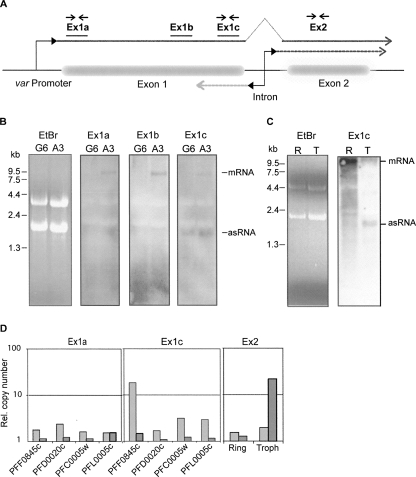
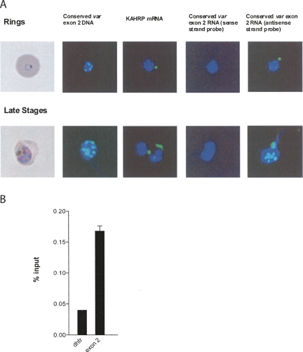
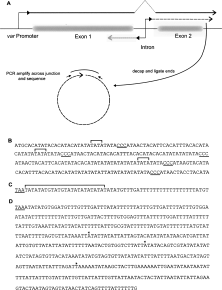
Antigenic variation by the malaria parasite Plasmodium falciparum results from switches in expression between members of the multicopy var gene family. These genes encode the variant surface protein PfEMP-1, the primary determinant of the antigenic and cytoadherent properties of infected erythrocytes. Only a single var gene is expressed at a time while the remaining members of the family remain transcriptionally silent. How mutually exclusive var gene expression is regulated is poorly understood; however, it is generally thought to involve alterations in chromatin assembly and modification, resulting in a type of cellular memory. Recently, several aspects of the chromatin structure surrounding var genes have been described, in particular the histone modifications associated with the active and silent states of the genes as well as their subnuclear localization. Here, we demonstrate that this chromatin structure also includes the incorporation of long sense and antisense noncoding RNAs. These sterile transcripts initiate from a bidirectional promoter located within a conserved intron found in all var genes that was previously implicated in var gene silencing. Mapping of the 5' and 3' ends of the sterile transcripts indicates that they are nonpolyadenylated. RNA fluorescent in situ hybridization (RNA-FISH) analysis detects both the sense and antisense noncoding RNAs in distinct spots within the nucleus similar to the pattern described for the var genes themselves. Further, analysis by RNA chromatin immunoprecipitation (ChIP) indicates that the noncoding RNAs are physically associated with chromatin. These sterile transcripts therefore might act in a manner analogous to noncoding RNAs associated with silent, condensed chromatin found in other eukaryotic systems.


Figures

FIGURE 1.
FIGURE 1.
Bidirectional promoter activity of var introns. Promoter activity of var introns was measured in transient transfection assays using the reporter construct pHLIRH (A), containing the reporter genes firefly and Renilla luciferase. Parasites were transfected and luciferase activity (firefly, light gray; Renilla dark gray) was measured from 2.5 mL cultures after ∼68 h (ring stage, light-colored columns) and ∼92 h (schizonts, dark columns), respectively (B). Luminescence values were standardized to 1% parasitemia. Genomic loci and upstream promoter types of var genes corresponding to the chosen var introns (grouping of var5′ promoter types according to Lavstsen et al. [2003]) (C).
FIGURE 2.
FIGURE 2.
Identification of exon 1 antisense RNA by Northern blot and quantitative RT-PCR. (A) The structure of a typical var gene is depicted schematically, and the relative positions of probes for Northern blot (solid lines) and PCR primer pairs (arrows) are indicated. Dashed lines represent the putative RNA transcripts originating in the intron in the sense (dark gray) and antisense (light gray) orientation. (B) Northern blot detection of RNA with specific probes for var gene PFF0845c. RNA was prepared from asynchronous cultures of NF54 clones A3 (PFF0845c “on”) and G6 (PFF0845c “off”). Major bands corresponding to var mRNA and antisense (as) transcripts are indicated. (C) Northern blot analysis using a PFF0845c exon 1 specific probe. RNA samples from synchronized A3 cultures were prepared at two different time points: in ring stage (R), ∼16 h after invasion and trophozoite stage (T), ∼36 h after invasion. For both B and C, ethidium bromide (EtBr) stained samples are included to show that approximately equal amounts of RNA were loaded in each lane. (D) var RNA transcripts in the transgenic DCJ line (Dzikowski et al. 2006) were detected by quantitative PCR using strand-specific cDNA as template created using either sense or antisense primers. Two primer pairs within exon 1 were specific for each of four var genes, while the primer pair within exon 2 recognized conserved sequences present in several var genes. The relative amounts of antisense and sense RNA, as determined by real-time PCR, are shown as light and dark gray columns, respectively. RNA was prepared from trophozoite stages (∼36 h post-invasion) of synchronous cultures, and for analysis with exon 2 primers additionally from ring stages (∼16 h).
FIGURE 3.
FIGURE 3.
Detection of var mRNA and antisense transcript by RNA-FISH. Single-stranded RNA probes were used to specifically detect mRNA (− strand) and exon 1 antisense transcript (+ strand) of var gene PFF0845c. A3 (PFF0845c “on”) and DCJ (PFF0845c “off”) parasites were analyzed 16–20 h post-invasion (ring stage), ∼30 hpi (trophozoite), and ∼40 hpi (schizont). Parasite nuclei were visualized by DAPI (blue). Overlays were generated of fluorescence pictures obtained by labeled RNA probes and DAPI stain, and one representative picture is shown for each sample.
FIGURE 4.
FIGURE 4.
Subcellular localization of the var sterile transcripts. (A) The clusters of var genes previously described can be seen in both rings and late trophozoites using DNA probes against exon 2. A larger number of clusters is seen in late trophozoites, indicating DNA replication has begun. RNA-FISH shows the localization of var transcripts in both rings (cytoplasmic, corresponding to the full-length, translated message) and the late trophozoites (nuclear, corresponding to the sterile transcripts). Sense strand RNA probes do not hybridize to either stage of parasites. The KAHRP RNA probe demonstrates cytoplasmic labeling as expected for a translated mRNA. (B) Chromatin immunprecipitation (ChIP) of var sterile transcripts with antibodies against histone H3 indicates that the sterile transcripts are associated with chromatin. Synchronized, late stage parasites were isolated and chromatin immunoprecipitated as described (Chookajorn et al. 2007). RNA was then isolated and cDNA analyzed using quantitative RT-PCR. The var sterile transcripts were more than fourfold more highly represented in the chromatin fraction than a representative mRNA dhfr. Precipitation with nonhistone antibodies (against PFI1780w) or experiments completed without reverse transcriptase did not give any signal.
FIGURE 5.
FIGURE 5.
Identification of the 5′ and 3′ ends of var transcripts. (A) Schematic diagram showing the RNA circularization method for simultaneous mapping of 5′ and 3′ ends. (B) The DNA sequence of a portion of the intron from var gene PFD0005w. The 5′ ends of the sterile RNAs transcribed from this gene are marked with brackets. The ccc repeat motif is underlined. (C) The DNA sequence downstream from the coding region of PFD0005w. The stop codon of the open reading frame is underlined. The sterile RNAs transcribed from this gene ended within 20 bp of the stop codon as marked by the bracket. (D) The DNA sequence downstream from the coding region of var gene PFD1005c. The polyadenylation sites of the mRNA transcribed from this gene are marked with asterisks. The stop codon is underlined.

References

    1. Akhtar A. Dosage compensation: An intertwined world of RNA and chromatin remodelling. Curr. Opin. Genet. Dev. 2003;13:161–169. - PubMed
    1. Andersson S.G.E., Zomorodipour A., Andersson J.O., Sicheritz-Ponten T., Alsmark U.C.M., Podowski R.M., Naslund A.K., Eriksson A.S., Winkler H.H., Kurland C.G. The genome sequence of Rickettsia prowazekii and the origin of mitochondria. Nature. 1998;396:133–140. - PubMed
    1. Aravind L., Iyer L.M., Wellems T.E., Miller L.H. Plasmodium biology: Genomic gleanings. Cell. 2003;115:771–785. - PubMed
    1. Balaji S., Babu M.M., Iyer L.M., Aravind L. Discovery of the principal specific transcription factors of Apicomplexa and their implication for the evolution of the AP2-integrase DNA binding domains. Nucleic Acids Res. 2005;33:3994–4006. - PMC - PubMed
    1. Baruch D.I., Pasloske B.L., Singh H.B., Bi X., Ma X.C., Feldman M., Taraschi T.F., Howard R.J. Cloning the P. falciparum gene encoding PfEMP1, a malarial variant antigen and adherence receptor on the surface of parasitized human erythrocytes. Cell. 1995;82:77–87. - PubMed

Publication types

MeSH terms

LinkOut - more resources