. 2009 Feb;20(3):769-79.
doi: 10.1091/mbc.e08-09-0903. Epub 2008 Nov 26.

Structural and functional requirements for activity of the Tim9-Tim10 complex in mitochondrial protein import

Affiliations

Structural and functional requirements for activity of the Tim9-Tim10 complex in mitochondrial protein import

Michael J Baker et al. Mol Biol Cell. 2009 Feb.

Abstract

The Tim9-Tim10 complex plays an essential role in mitochondrial protein import by chaperoning select hydrophobic precursor proteins across the intermembrane space. How the complex interacts with precursors is not clear, although it has been proposed that Tim10 acts in substrate recognition, whereas Tim9 acts in complex stabilization. In this study, we report the structure of the yeast Tim9-Tim10 hexameric assembly determined to 2.5 A and have performed mutational analysis in yeast to evaluate the specific roles of Tim9 and Tim10. Like the human counterparts, each Tim9 and Tim10 subunit contains a central loop flanked by disulfide bonds that separate two extended N- and C-terminal tentacle-like helices. Buried salt-bridges between highly conserved lysine and glutamate residues connect alternating subunits. Mutation of these residues destabilizes the complex, causes defective import of precursor substrates, and results in yeast growth defects. Truncation analysis revealed that in the absence of the N-terminal region of Tim9, the hexameric complex is no longer able to efficiently trap incoming substrates even though contacts with Tim10 are still made. We conclude that Tim9 plays an important functional role that includes facilitating the initial steps in translocating precursor substrates into the intermembrane space.


Figures

Figure 1.
Figure 1.
Crystal structure of the yeast Tim9–Tim10 hexameric complex. (A) Ribbon diagram of the Tim9–Tim10 complex with a side view (left) and an aerial view (right). Tim9 is in red and Tim10 is in blue. (B) Ribbon diagrams depicting the individual Tim9 and Tim10 subunits forming hairpin-like structures braced by two intramolecular disulfide bonds (yellow). (C) Ribbon diagram depicting the interaction between Met25 of Tim9 and Leu28 of Tim10. (D) Crystal lattice packing of the Tim9–Tim10 complex. Residues are colored according to their individual B-factor. Blue corresponds to residues with low thermal mobility and red corresponds to residues with high thermal mobility. The B-factors ranges from 35 to 153 Å2. (E) -Fold comparison of the human and yeast subunits within the Tim9–Tim10 complex. hTim9 (orange) is superimposed on yTim9 (red), and hTim10 (green) has been superimposed on yTim10 (blue). (F) Ribbon diagram depicting Tyr49 of Tim10 packing into the core of an adjacent Tim9–Tim10 complex.
Figure 2.
Figure 2.
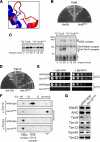
Disruption of a conserved salt-bridge between Tim9 and Tim10. (A) Ribbon diagram showing the formation of the Tim9E52–Tim10K68 salt bridge. (B) Growth of yeast strains expressing wild-type Tim9, Tim9E52K, or lacking Tim9 plated on minimal glucose media supplemented with 5-FOA at 24°C. (C) 35S-Tim9 and 35S-Tim9E52K were imported into yeast mitochondria at 24°C for the times indicated. Mitochondria were reisolated and subjected to SDS-PAGE (left) or BN-PAGE (right) followed by phosphorimaging (*, large Tim9–Mia40 complex; #, Tim9–Tim12 intermediate complex; u, unassembled protein). (D) Growth of yeast strains expressing wild-type Tim10, Tim10K68E, or lacking Tim10 plated on minimal glucose media supplemented with 5-FOA at 24°C. (E) Serial dilutions of yeast strains expressing wild-type Tim10 or Tim10K68E were generated and spotted onto nonfermentable (+ glycerol) or fermentable (+ glucose) media and incubated at 24°C or 37°C as indicated. (F) Mitochondria from yeast strains expressing wild-type Tim10 or Tim10K68E were solubilized and run on BN-PAGE in the first dimension and SDS-PAGE in the second dimension before Western blot analysis (*, nonassembled protein). (G) Equal amounts of wild-type or tim10K68E mitochondria were subjected to SDS-PAGE and subsequent Western blot analysis using marker antibodies, as indicated.
Figure 3.
Figure 3.
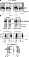
Analysis of protein import in tim10K68E mitochondria. (A) 35S-AAC was imported into wild-type or tim10K68E mitochondria preincubated at 24°C (− heat shock) or 37°C (+ heat-shock) before BN-PAGE analysis. Dissipation of the Δψ blocks AAC insertion into the inner membrane (AACII, AAC dimer). (B) 35S-Tom40 was imported into wild-type and tim10K68E mitochondria preincubated at 24°C (− heat shock) or 37°C (+ heat shock). Tom40 assembly intermediates were analyzed by BN-PAGE and phosphorimaging (SAM intermediate, Tom40 precursor at the SAM complex; intermediate II, Tom40 intermediate integrated into the outer membrane). (C) Wild-type or tim10K68E mitochondria were either pretreated with a heat shock or not subjected to a heat shock before incubation with 35S-AAC-DHFR in the presence of methotrexate. After cross-linking, mitochondria were lysed and subjected to immunoprecipitation with antibodies against Tim9, Tim10, and Tom40 before SDS-PAGE and phosphorimaging. Some AAC-DHFR is nonspecifically pulled down (Tom40-XL, cross-links formed between Tom40 and AAC-DHFR; arrow, Tim9 or Tim10 cross-links with AAC-DHFR). (D) 35S-AAC-DHFR arrested at the TOM complex in wild-type and tim10K68E mitochondria was analyzed using BN-PAGE and phosphorimaging (left). Level of arrested AAC-DHFR was quantified (right; error bars represent SD; n = 3).
Figure 4.
Figure 4.
Generation of Tim9 and Tim10 truncation mutants. (A) Schematic representation of Tim10 truncation mutants. Arrows point to the most N- or C-terminal residue remaining in the truncation mutants. Growth phenotypes are noted. (B) The growth of yeast strains expressing Tim10 and truncation mutants was analyzed at 24 or 37°C by serially diluting and spotting yeast onto glucose or glycerol containing media. (C) Schematic representation of Tim9 truncation mutants. Arrows point to the most N- or C-terminal residue remaining in the truncation mutants. Growth phenotypes are noted. (D) The growth of yeast strains expressing Tim9 and truncation mutants was analyzed at 24 or 37°C by serially diluting and spotting yeast onto glucose- or glycerol-containing media.
Figure 5.
Figure 5.
Analysis of TIM22 and Tim9–Tim10 complexes in truncation mutants. (A–D) Mitochondria isolated from the yeast strains indicated were subjected to BN-PAGE before SDS-PAGE in the second dimension followed by Western blot analysis. The positions of the TIM22 and Tim9–Tim10 complexes are indicated. (*, nonassembled forms of the proteins).
Figure 6.
Figure 6.
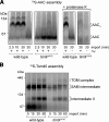
Import analysis in tim9ΔN10 mitochondria. (A) 35S-AAC was imported into wild-type and tim9ΔN10 mitochondria (without a heat-shock pretreatment) at 24°C in the presence and absence of Δψ. After treating some samples to proteinase K, mitochondria were isolated, solubilized, and analyzed using BN-PAGE and phosphorimaging. Right, AAC complexes (AAC and AACII) protected from proteinase K treatment of intact mitochondria. (B) 35S-Tom40 was imported into wild-type and tim9ΔN10 mitochondria (without heat shock) at 24°C and analyzed using BN-PAGE and phosphorimaging. (SAM intermediate, Tom40 precursor at the SAM complex; intermediate II, Tom40 intermediate integrated into the outer membrane).
Figure 7.
Figure 7.
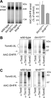
Accumulation and cross-linking analysis of AAC-DHFR at the TOM complex. (A) 35S-AAC-DHFR was incubated with mitochondria isolated from indicated yeast strains at 24°C in the presence of methotrexate and then analyzed using BN-PAGE and phosphorimaging (left). The levels of TOM-arrested AAC-DHFR were quantified (right; error bars represent SD, n = 3). (B) 35S-AAC-DHFR was incubated with wild-type or tim10ΔN12 mitochondria in the presence of methotrexate. Cross-linking to Tim9, Tim10ΔN12, and Tom40 was assessed by immunoprecipitation and SDS-PAGE (Tom40-XL, cross-links formed between Tom40 and AAC-DHFR; arrow, cross-links formed between either Tim9 or Tim10 and AAC-DHFR). Some noncross-linked AAC-DHFR is nonspecifically pulled down. (C) 35S-AAC-DHFR was incubated with tim9ΔN10 mitochondria in the presence of methotrexate and subjected to cross-linking analysis as described in B.

Similar articles

Cited by

References

    1. Allen S., Balabanidou V., Sideris D. P., Lisowsky T., Tokatlidis K. Erv1 mediates the Mia40-dependent protein import pathway and provides a functional link to the respiratory chain by shuttling electrons to cytochrome c. J. Mol. Biol. 2005;353:937–944. - PubMed
    1. Baker M. J., Frazier A. E., Gulbis J. M., Ryan M. T. Mitochondrial protein-import machinery: correlating structure with function. Trends Cell Biol. 2007;17:456–464. - PubMed
    1. Bolender N., Sickmann A., Wagner R., Meisinger C., Pfanner N. Multiple pathways for sorting mitochondrial precursor proteins. EMBO Rep. 2008;9:42–49. - PMC - PubMed
    1. Brunger A. T., et al. Crystallography & NMR system: a new software suite for macromolecular structure determination. Acta Crystallogr. D Biol. Crystallogr. 1998;54:905–921. - PubMed
    1. Chacinska A., et al. Mitochondrial presequence translocase: switching between TOM tethering and motor recruitment involves Tim21 and Tim17. Cell. 2005;120:817–829. - PubMed

Publication types

MeSH terms

Substances

LinkOut - more resources