. 2009 Feb;20(3):846-58.
doi: 10.1091/mbc.e08-08-0830. Epub 2008 Nov 26.

Fibrillin assembly requires fibronectin

Affiliations

Fibrillin assembly requires fibronectin

Laetitia Sabatier et al. Mol Biol Cell. 2009 Feb.

Abstract

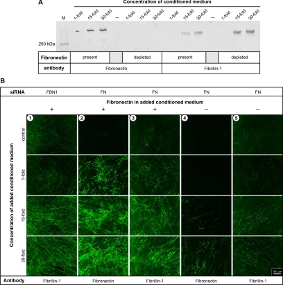
Fibrillins constitute the major backbone of multifunctional microfibrils in elastic and nonelastic extracellular matrices. Proper assembly mechanisms are central to the formation and function of these microfibrils, and their properties are often compromised in pathological circumstances such as in Marfan syndrome and in other fibrillinopathies. Here, we have used human dermal fibroblasts to analyze the assembly of fibrillin-1 in dependence of other matrix-forming proteins. siRNA knockdown experiments demonstrated that the assembly of fibrillin-1 is strictly dependent on the presence of extracellular fibronectin fibrils. Immunolabeling performed at the light and electron microscopic level showed colocalization of fibrillin-1 with fibronectin fibrils at the early stages of the assembly process. Protein-binding assays demonstrated interactions of fibronectin with a C-terminal region of fibrillin-1, -2, and -3 and with an N-terminal region of fibrillin-1. The C-terminal half of fibrillin-2 and -3 had propensities to multimerize, as has been previously shown for fibrillin-1. The C-terminal of all three fibrillins interacted strongly with fibronectin as multimers, but not as monomers. Mapping studies revealed that the major binding interaction between fibrillins and fibronectin involves the collagen/gelatin-binding region between domains FNI(6) and FNI(9).


Figures

Figure 1.
Figure 1.
Expression and network formation of the fibril-forming proteins fibronectin and fibrillin-1 after siRNA treatment. Human dermal fibroblasts were treated with siRNA oligonucleotides for fibronectin (FN) or fibrillin-1 (FBN1) as described in Materials and Methods and grown for a predetermined optimum time for each application (4 d for RNA extraction; 2 d for FN staining; 4 d for FBN1 staining). No effect on fibronectin or fibrillin-1 assembly was observed with control siRNAs (Allstars). (A) Fibronectin and fibrillin-1 network formation was monitored by indirect immunofluorescence as indicated. All images had a similar cell density as controlled by nuclear DAPI staining. Bar, 100 μm for all images. (B) The total RNA was extracted, reverse transcribed and analyzed by real-time PCR. The mRNA expression was normalized to the expression of GAPDH. Signals from the AllStars siRNA negative control were set to 100%. (C) Conditioned medium (0.33 ml) was analyzed by Western blotting using the specific antibodies indicated. All bands migrated at positions characteristic for each protein above the highest marker protein (250 kDa) used.
Figure 2.
Figure 2.
Network formation of fibrillin-1 in the presence of bacterially derived fibronectin-binding peptides. (A) Shown is an indirect immunofluorescence after seeding human dermal fibroblasts with either no treatment (−), or peptide FUD, which inhibits fibronectin network formation, or peptide Del29, which is an inactive mutant of FUD. The peptides were added at a concentration of 500 nM. Note that FUD completely blocks fibronectin assembly and also fibrillin-1 assembly, but the Del29 peptide had no influence on both. DAPI staining confirmed equal numbers of cells in all images. Bar, 50 μm for all images. (B) As a control, fibroblasts were treated in the presence of the indicated peptides identically as in A, except that the medium was replaced by serum-free (peptide-containing) medium for the 24-h condition period. Shown is a Western blot analysis of 1 ml conditioned culture medium stained with specific antibodies against fibronectin and fibrillin-1 as indicated.
Figure 3.
Figure 3.
Network formation by exogenous fibronectin promotes assembly of exogenous fibrillin-1. Conditioned fibroblast culture medium, with or without fibronectin, was produced and concentrated as described in Materials and Methods. (A) Western blot analysis of the conditioned medium with antibodies against fibronectin or fibrillin-1 shows the efficacy of the fibronectin depletion, whereas the level of fibrillin-1 was not reduced. (B) Fibroblasts were treated with siRNA to reduce the level of fibrillin-1 (FBN1) or fibronectin (FN) expression. The conditioned media with (+) or without (−) fibronectin as analyzed in A were added to the cultures for 48 h, and the formation of fibrillin-1 and fibronectin networks were visualized by indirect immunofluorescence using antibodies against these proteins. Similar results were obtained when the cell cultures were incubated for 20 h with the conditioned media. The presence of similar numbers of cells in all images was verified with DAPI staining of cell nuclei. Additional sets of images for column 1 are shown in Supplemental Figure S1. Bar, 100 μm for all images.
Figure 4.
Figure 4.
Fibrillin-1 and fibronectin colocalize in the matrix produced by dermal fibroblasts. (A) Shown is indirect immunofluorescence labeling of networks which are assembled either from endogenously produced (row 1) or from exogenously added (row 2) fibrillin-1 and fibronectin. The cells in row 1 were not treated with siRNA, allowing endogenously produced fibronectin and fibrillin-1 to assemble. Cells in rows 2 and 3 were treated with siRNAs for both FBN1 and FN to reduce simultaneously the endogenous expression of fibrillin-1 and fibronectin. Conditioned medium containing fibrillin-1 and fibronectin (15-fold concentrated), as characterized in Figure 3A, was added to cells in row 2 and nonconditioned control medium was added to cells in row 3 as described in Materials and Methods. DAPI staining of cell nuclei verified similar cell densities in each field. Bar, 100 μm for all images. (B) Shown is an ELISA analysis to demonstrate that cross-reactivity of the antibodies for fibrillin-1 and fibronectin is negligible. The polyclonal antiserum α-rFBN1-C (red and blue symbols) and the mAb FN-15 (green, yellow, and white symbols) were tested with rFBN1-N (downward-pointing triangles) and rFBN1-C (circles and upward-pointing triangles) or full-length fibronectin (FN; diamonds and squares) as indicated. (C) Shown is a double-immunogold localization of extracellular fibrils produced by human dermal fibroblasts after 3 d in culture. Eighteen-nanometer gold particles represent fibrillin-1, and 12-nm gold particles represent fibronectin. Bar, 100 nm for all images.
Figure 5.
Figure 5.
Schematic overview of recombinant fibrillin and fibronectin fragments used in this study. The N-terminal half of fibrillin-3 was not available as a recombinant purified protein. The proteins have been analyzed under nonreducing (−) and reducing (+) conditions using DTT (insets). Molecular marker proteins are indicated in kDa.
Figure 6.
Figure 6.
Interactions of fibrillins with fibronectin. Shown is a representative solid-phase binding assay with coated human plasma fibronectin and the soluble halves of fibrillin-1, -2, and -3 in concentrations as indicated. Solid symbols represent binding in the presence of 5 mM CaCl2, and open symbols represent binding of calcium-free fibrillin fragments in the presence of 10 mM EDTA. Note that the C-terminal half of all fibrillins bound dose-dependently and calcium-independently to fibronectin. The N-terminal half of fibrillin-1 bound moderately to fibronectin in the presence of calcium. A control interaction of rFBN1-C with rFBN1-N, known to be calcium-dependent (Lin et al., 2002), showed full calcium dependency under identical assay conditions (not shown). Data represent means of duplicates; error bars, SDs.
Figure 7.
Figure 7.
Multimerization of the fibrillin-1, -2, and -3 C-terminal half into large-molecular-weight assemblies and interaction of monomers and multimers with fibronectin. (A) The recombinant C-terminal half of fibrillin-1 (rFBN1-C) as an established control (Hubmacher et al., 2008), fibrillin-2 (rFBN2-C), and fibrillin-3 (rFBN3-C) were subjected to gel filtration chromatography (top panel). Plotted are the elution volumes versus their absorbance at 280 nm (mA units). Aliquots (20 μl) from the elution volumes indicated were analyzed by silver staining after SDS gel electrophoresis under reducing (+) and nonreducing (−) conditions (DTT; bottom panel). Molecular marker proteins are indicated in kDa. The positions of multimeric (Mu), intermediate (In), and monomeric (Mo) fragments are indicated in each panel. Note that both, fibrillin-2 and -3 C-terminal fragments form reducible high-molecular-weight assemblies similar to fibrillin-1, albeit with some variations in the total amounts. The position of the reducible fibrillin monomers in the gels are indicated by arrowheads. (B) Solid-phase interaction assay with soluble rFBN1-C, rFBN2-C, and rFBN3-C (as indicated) in the monomeric (○) and multimeric (□) states with immobilized full-length plasma fibronectin. Data represent means of duplicates; error bars, SDs. Note that only the fibrillin multimers bind to fibronectin but not the monomers.
Figure 8.
Figure 8.
Mapping of fibrillin binding sites on fibronectin. Shown are representative solid phase interaction assays. The recombinant fibrillin-1, -2, and -3 C-termini (A) or fibrillin-1 and -2 N-termini (B) were analyzed as soluble ligands with immobilized recombinant fibronectin fragments FNsuper70K (○) or FNIII1-C (□), or with proteolytic fragment FN40K (▿). Efficient immobilization of fibronectin fragments was verified by ELISA assays (see Supplemental Figure S3). Data points represent means of duplicates; error bars, SDs.
Figure 9.
Figure 9.
Inhibition of fibrillin–fibronectin interactions by gelatin. Gelatin was first bound in increasing concentrations (A) to immobilized full-length plasma fibronectin and (B) to the fibronectin fragments FNsuper70K and FN40K. In a second incubation step at constant ligand concentrations, fibrillin-1, -2, and -3 C-terminal halves (as indicated) were bound to the immobilized full-length fibronectin (A), or the fibrillin-1 N-terminal half was bound to the immobilized fibronectin fragments (B). The signal without gelatin was set to 100%. Data points represent means of duplicates; error bars, SDs.
Figure 10.
Figure 10.
Binding of fluorescence-labeled recombinant halves of fibrillin-1 to matrix produced by dermal fibroblasts. Fragments rFBN1-N and rFBN1-C were covalently labeled with the fluorescent dye Cy3 and then added at 10 μg/ml (60 or 70 nM, respectively) for 24 h to the culture medium of cells precultured for 24 h. The cells were then washed and labeled with an mAb against fibronectin followed by FITC-conjugated secondary antibody. Shown is the fluorescence microscopic analysis of the localization of the Cy3-labeled fibrillin fragments (red) and the fibronectin staining (green). Note that the Cy3-labeled fibrillin fragments colocalize with fibronectin labeled fibrils. DAPI staining shows the density and distribution of the cell nuclei in the image fields. Bar, 100 μm for all images.

Similar articles

Cited by

References

    1. Akiyama S. K., Yamada S. S., Chen W. T., Yamada K. M. Analysis of fibronectin receptor function with monoclonal antibodies: roles in cell adhesion, migration, matrix assembly, and cytoskeletal organization. J. Cell Biol. 1989;109:863–875. - PMC - PubMed
    1. Ashworth J. L., Kelly V., Wilson R., Shuttleworth C. A., Kielty C. M. Fibrillin assembly: dimer formation mediated by amino-terminal sequences. J. Cell Sci. 1999;112:3549–3558. - PubMed
    1. Baldock C., Koster A. J., Ziese U., Rock M. J., Sherratt M. J., Kadler K. E., Shuttleworth C. A., Kielty C. M. The supramolecular organization of fibrillin-rich microfibrils. J. Cell Biol. 2001;152:1045–1056. - PMC - PubMed
    1. Baldock C., et al. Nanostructure of fibrillin-1 reveals compact conformation of EGF arrays and mechanism for extensibility. Proc. Natl. Acad. Sci. USA. 2006;103:11922–11927. - PMC - PubMed
    1. Balian G., Click E. M., Crouch E., Davidson J. M., Bornstein P. Isolation of a collagen-binding fragment from fibronectin and cold-insoluble globulin. J. Biol. Chem. 1979;254:1429–1432. - PubMed

Publication types

MeSH terms

LinkOut - more resources