. 2009 Feb;20(3):780-90.
doi: 10.1091/mbc.e08-03-0256. Epub 2008 Nov 26.

Early stages of Golgi vesicle and tubule formation require diacylglycerol

Affiliations

Early stages of Golgi vesicle and tubule formation require diacylglycerol

Lennart Asp et al. Mol Biol Cell. 2009 Feb.

Abstract

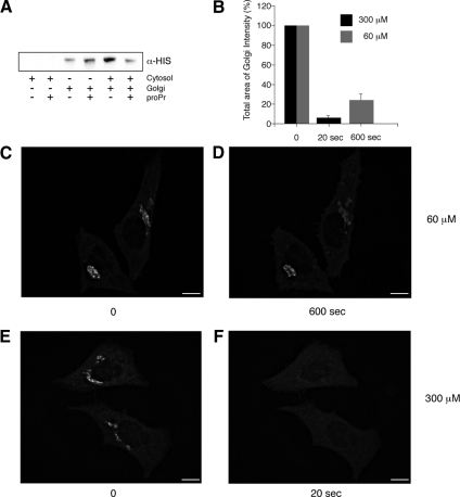
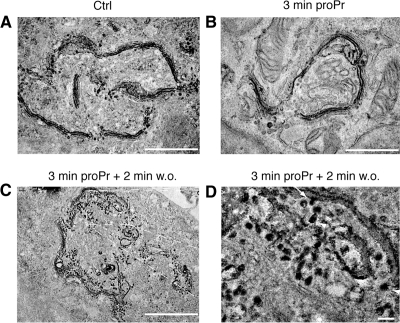
We have investigated the role for diacylglycerol (DAG) in membrane bud formation in the Golgi apparatus. Addition of propranolol to specifically inhibit phosphatidate phosphohydrolase (PAP), an enzyme responsible for converting phosphatidic acid into DAG, effectively prevents formation of membrane buds. The effect of PAP inhibition on Golgi membranes is rapid and occurs within 3 min. Removal of the PAP inhibitor then results in a rapid burst of buds, vesicles, and tubules that peaks within 2 min. The inability to form buds in the presence of propranolol does not appear to be correlated with a loss of ARFGAP1 from Golgi membranes, as knockdown of ARFGAP1 by RNA interference has little or no effect on actual bud formation. Rather, knockdown of ARFGAP1 results in an increase in membrane buds and a decrease of vesicles and tubules suggesting it functions in the late stages of scission. How DAG promotes bud formation is discussed.


Figures

Figure 1.
Figure 1.
Inhibition of DAG formation through PAP1 results in a rapid loss of ARFGAP1 from Golgi membranes. (A) Inhibition of ARFGAP1 binding to Golgi membranes by proPr is cytosol-dependent. Recombinant His-tagged ARFGAP1 (0.1 μg) was incubated in 200 μl reaction buffer (see Material and Methods) together with highly purified Golgi membranes (20 μg), rat liver cytosol (1 mg), or proPr (300 μM) for 15 min at 37°C. Incubation mixtures were terminated on ice and membranes pelleted at 13 000 rpm for 10 min. Solubilized proteins were then separated by SDS-PAGE and transferred to nitrocellulose for Western blotting. His-ARFGAP1 was detected using a poly-His specific antibody followed by secondary HRP-labeled rabbit anti-mouse antibody and an ECL detection system. (B–F) Addition of 60 μM proPr for 600 s (C and D) or 300 μM proPr for 20 s in HeLa cells expressing ARFGAP1EGFP (E and F), respectively, results in a partial or complete loss of the Golgi-localized ARFGAP1EGFP. (B) Quantitation (abscissa) as described in Materials and Methods. Scale bars, (C–F) 10 μm.
Figure 2.
Figure 2.
DAG is required for bud formation. Thin plastic embedded sections (60 nm thick) of HeLa cells were examined at the ultrastructural level. (A–C) Representative low-magnification fields (scale bar, 1 μm), whereas D–G show representative high-magnification fields (scale bar, 100 nm). (A and D) Multiple Golgi stacks align laterally to form a part of the Golgi ribbon in untreated cells. Associated membrane buds (arrow) and VTPs (arrowhead) were seen in close proximity to cisternal membranes of the Golgi stack. (B and E) Addition of 300 μM proPr for 3 min resulted in an increased frequency of curved stacks that consisted of smooth cisternal membranes seemingly devoid of both membrane buds as well as VTPs. Occasional VTPs and buds (arrowhead in E) were observed but at a marked decreased frequency (see G for quantitation). In C and F, removal of proPr resulted in a dramatic increase of both membrane buds as well as VTPs already after 2 min. Arrow and arrowheads point to a bud and VTPs, respectively. (G) Quantitation of cisternae, VTPs, and membrane buds presented as the mean of total membranes and compared with untreated control (Ctrl), which was set at 100%.
Figure 3.
Figure 3.
Electron tomography of Golgi stacks. Dual axis tomography was performed to obtain good resolution of membrane delineations in all specimen planes. (A) A digital slice through the 3D volume of an electron tomographic reconstruction illustrates the appearance of the Golgi area seen before addition of proPr (left field), 3 min after addition of proPr (middle field) and 2 min after removal of proPr (right field). Each membrane-delineated structure present in this digital slice was analyzed throughout the 3D volume and color-coded. Blue, Golgi cisterna or structures continuous with a Golgi cisterna except membrane buds; red, vesicles; green, tubular structures; and yellow, membrane buds. Scale bar, 100 nm. (B) Different close-up fields vesicles and membrane buds observed after removal of proPr. Arrows indicate necks of budding profiles that appear constricted by electron-dense material. Scale bar, 40 nm.
Figure 4.
Figure 4.
Inhibition of DAG formation prevents GalNAcT2CFP from entering VTPs. After photooxidation and epon embedding, DAB precipitate was examined by electron microscopy. (A) DAB precipitate is predominantly found in 2–3 cisternae reflecting a gradient-like distribution across the Golgi stack. Some associated VTPs are also positive for the DAB precipitate, consistent with that GalNAcT2CFP can gain access to these structures. (B) On addition of proPr (300 μM) for 3 min, the DAB precipitate is seen exclusively in cisternal membranes but cannot be detected in any associated buds or VTPs. (C) At 2 min after removal of proPr, the DAB product is seen in both cisternal membranes as well as VTPs. (D) Magnified field corresponding to the box in C. Arrow points to a bud-like structure filled with DAB precipitate. Arrowheads point to VTPs with a diameter of 40–50 nm. Scale bar, (A–C) 1 μm; (D) 75 nm.
Figure 5.
Figure 5.
ARFGAP1 is required for membrane fission. Thin plastic embedded sections (60 nm thick) of HeLa cells transfected with either RNAiMock or RNAiARFGAP1 were examined at the ultrastructural level to discern structures associated with Golgi stacks. Observed structures were quantified in D as in Figure 2. (A and D) RNAiARFGAP1-transfected cells revealed an increased frequency of membrane buds accompanied by a decreased number of associated VTPs compared with RNAiMock-transfected cells. (B and D) Addition of proPr (300 μM) for 3 min resulted in a marked decrease in associated membrane buds and VTPs in RNAiARFGAP1-transfected cells. (C and D) Removal of proPr revealed a marked increase in membrane buds in cells transfected with RNAiARFGAP1 compared with cells transfected with RNAiMock over that of associated VTPs. Scale bar, 100 nm.
Figure 6.
Figure 6.
Schematic overview and model. (A) Inhibition of DAG synthesis in the Golgi results in an inhibition of bud formation after incubation with 300 μM proPr for 3 min. Observed Golgi stacks lack definable bud structures as well as adjacent vesicles and tubules and instead appear smooth and nonfenestrated compared with untreated cells. Resumption of DAG synthesis results in a rapid increase in bud, vesicle, and tubular formation already after 2 min. At 5–10 min after washout, stacks have reformed. This reveals an underlying rapid and dynamic behavior of Golgi membranes in response to DAG synthesis. Knockdown of ARFGAP1 through siRNA experiments results in Golgi stacks with associated buds but no adjacent vesicles and tubules. In such cells, addition of proPr for 3 min results in Golgi stacks devoid of buds. Taken together, this suggests a role for DAG in bud formation and a role for ARFGAP1 in later stages of vesicle and tubule formation (e.g., to promote vesicle scission). (B) A model illustrates the proposed function of DAG in terms of promoting bud formation. (1) COPI coat-components such as coatomer, ARF1, and ARFGAP1 bind to the cytosolic leaflet of the Golgi-cisternal membrane. (2a) Formation of LPA (an inverted cone-shaped lipid, depicted in blue) and DAG (a cone-shaped lipid, depicted in red), allows the membrane to bend outward (2b) into the cytosol. The ability of DAG to rapidly flip-flop (FF) over to the luminal leaflet explains why DAG is required for bud formation in its ability to promote negative curvature. (3) The bud has formed with LPA enriched in the cytosolic leaflet at the tip with DAG in the opposite luminal leaflet. At the base of the bud, PA- and DAG-binding proteins are proposed to help stabilize the intermediate. As DAG can easily flip-flop back to the cytosolic leaflet, it has been suggested that its presence causes disorder in the membrane and as a consequent results in an increased ability of ARFGAP1 to promote GTP-hydrolysis of ARF1 resulting in a loss of coatomer once the vesicle has been formed and released (scission). Note that LPA can be converted back into PA and used to form DAG. Equally well, DAG can be converted back to PA, which then can be used to for LPA. How this is regulated to promote COPI vesicle formation is presently unknown. GPL, general phospholipid.

References

    1. Allan V. J., Kreis T. E. A microtubule-binding protein associated with membranes of the Golgi apparatus. J. Cell Biol. 1986;103:2229–2239. - PMC - PubMed
    1. Antonny B., Bigay J., Casella J. F., Drin G., Mesmin B., Gounon P. Membrane curvature and the control of GTP hydrolysis in Arf1 during COPI vesicle formation. Biochem. Soc. Trans. 2005;33:619–622. - PubMed
    1. Antonny B., Huber I., Paris S., Chabre M., Cassel D. Activation of ADP-ribosylation factor 1 GTPase-activating protein by phosphatidylcholine-derived diacylglycerols. J. Biol. Chem. 1997;272:30848–30851. - PubMed
    1. Aoe T., Cukierman E., Lee A., Cassel D., Peters P. J., Hsu V. W. The KDEL receptor, ERD2, regulates intracellular traffic by recruiting a GTPase-activating protein for ARF1. EMBO J. 1997;16:7305–7316. - PMC - PubMed
    1. Bai J., Pagano R. E. Measurement of spontaneous transfer and transbilayer movement of BODIPY-labeled lipids in lipid vesicles. Biochemistry. 1997;36:8840–8848. - PubMed