. 2009 Jan 15;457(7227):318-21.
doi: 10.1038/nature07472. Epub 2008 Nov 26.

Role for Spi-C in the development of red pulp macrophages and splenic iron homeostasis

Affiliations

Role for Spi-C in the development of red pulp macrophages and splenic iron homeostasis

Masako Kohyama et al. Nature. .

Abstract

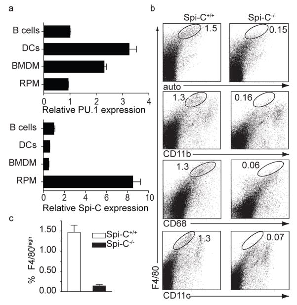
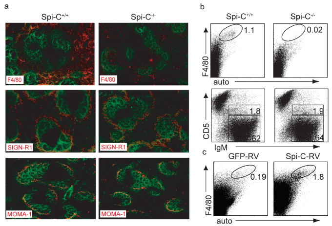
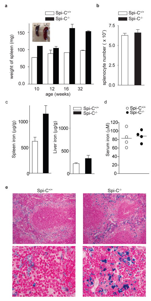
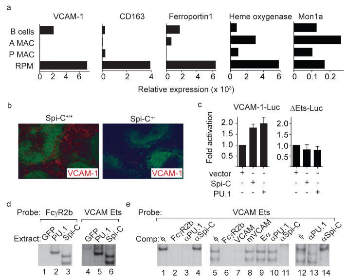
Tissue macrophages comprise a heterogeneous group of cell types differing in location, surface markers and function. Red pulp macrophages are a distinct splenic subset involved in removing senescent red blood cells. Transcription factors such as PU.1 (also known as Sfpi1) and C/EBPalpha (Cebpa) have general roles in myelomonocytic development, but the transcriptional basis for producing tissue macrophage subsets remains unknown. Here we show that Spi-C (encoded by Spic), a PU.1-related transcription factor, selectively controls the development of red pulp macrophages. Spi-C is highly expressed in red pulp macrophages, but not monocytes, dendritic cells or other tissue macrophages. Spic(-/-) mice have a cell-autonomous defect in the development of red pulp macrophages that is corrected by retroviral Spi-C expression in bone marrow cells, but have normal monocyte and other macrophage subsets. Red pulp macrophages highly express genes involved in capturing circulating haemoglobin and in iron regulation. Spic(-/-) mice show normal trapping of red blood cells in the spleen, but fail to phagocytose these red blood cells efficiently, and develop an iron overload localized selectively to splenic red pulp. Thus, Spi-C controls development of red pulp macrophages required for red blood cell recycling and iron homeostasis.


Figures

Figure 1
Figure 1. Spi-C−/− mice have a selective loss of red pulp macrophages
a, PU.1 and Spi-C eexpression was determined by quantitative RT-PCR in purified B cells, dendritic cells (DCs), BM-derived macrophages (BMDM), and red pulp macrophages (RPM). Shown is the normalized mRNA expression relative to expression in B cells. b, Spi-C+/+ and Spi-C−/− spleen cells were stained with antibodies to F4/80, CD11b, CD68, and CD11c and analyzed by flow cytometry. Numbers represent the percentage of cells in the indicated gate. c, Frequency of F4/80hi cells in spleen was determined as a mean (± SD) (n=7) from total splenocytes as shown in b.
Figure 2
Figure 2. Spi-C−/− mice have a cell-autonomous defect in red pulp macrophages
a, Spi-C+/+ and Spi-C−/− spleens sections were stained for B220 (green) and F4/80, SIGN-R1 or MOMA-1 (red). b, BM cells from CD45.2+ C57BL/6 Spi-C+/+ or Spi-C−/− mice were transferred into irradiated CD45.1+ B6.SJL mice. Splenocytes were stained for CD45.2, CD45.1, F4/80, CD5 and IgM. After 10 weeks, >97% of spleen cells were donor-derived (CD45.2+CD45.1). Plots are gated on donor-derived cells. Numbers represent the percentage of donor-derived cells in the indicated gates. c, BM cells from CD45.2+ C57BL/6 Spi-C−/− mice were infected with Spi-C-RV or control RV and transferred into irradiated CD45.1+ B6.SJL mice. After 6 weeks, spleen cells were stained for CD45.2, CD45.1, and F4/80. Plots are gated on donor-derived cells. Results are representative of four mice per group.
Figure 3
Figure 3. Spi-C−/− mice have increased splenic iron stores
a, Spleens from Spi-C+/+ and Spi-C−/− mice of the indicated age were weighed (n=3 per time point). Representative spleens at 32 weeks (Inset). b, Splenocytes from Spi-C+/+ and Spi-C−/− mice were counted (mean ± SD, n=5). c, Iron levels in spleen (left) and liver (right) of 129 SvEv Spi-C+/+ and Spi-C−/− male mice on a standard diet (mean ± SD, n=4). d, Serum iron levels were determined for 32 weeks old 129 SvEv background Spi-C+/+ and Spi-C−/− male mice on a standard diet. e, Perl’s Prussian blue stain for ferric iron in the spleens of 16-week old 129 SvEv Spi-C+/+ and Spi-C−/− male mice.
Figure 4
Figure 4. Spi-C regulates VCAM-1 expression
a, Normalized expression for VCAM-1, CD163, ferroportin I, heme oxygenase, and Mon1a are shown for B cells, alveolar macrophages (A MAC), peritoneal macrophages (P MAC) and red pulp macrophages (RPM) as described in Fig. 1. b, Spi-C+/+ or Spi-C−/− spleen sections were stained for B220 (green) and VCAM-1 (red). c, J774 cells were transfected with the VCAM-1 reporter (VCAM-1-Luc) or the ΔEts-Luc reporter, and with pEF4 (vector), Spi-C- or PU.1-expressing vectors. Cells were analyzed for luciferase activity as described in the Methods. Data is representative of 4 independent experiments (mean ± SD, n=3). d, 293F/T cells were transiently transfected with GFP-RV (GFP), PU1-MIGR1 (PU.1) or Spi-C-RV (Spi-C) and whole cell extracts were analyzed for binding to the FCγR2b or the VCAM Ets probes. e, Extracts from PU.1-expressing cells (lanes 1–4), Spi-C-expressing cells (lanes 5–11) or mixtures of PU.1 and Spi-C extracts (lanes 12–14) were analyzed for binding to the VCAM Ets probe. Shown are competitions (Comp) using unlabelled competitor oligonucleotides or supershifts using anti-sera against PU.1 or Spi-C as indicated.

References

    1. Taylor PR, et al. Macrophage receptors and immune recognition. Ann Rev Immunol. 2005;23:901–944. - PubMed
    1. Gordon S, Taylor PR. Monocyte and macrophage heterogeneity. Nature Reviews Immunology. 2005;5:953–964. - PubMed
    1. Ye M, Graf T. Early decisions in lymphoid development. Curr Opin Immunol. 2007;19:123–128. - PubMed
    1. Friedman AD. Transcriptional control of granulocyte and monocyte development. Oncogene. 2007;26:6816–6828. - PubMed
    1. Bemark M, Martensson A, Liberg D, Leanderson T. Spi-C, a novel Ets protein that is temporally regulated during B lymphocyte development. J Biol Chem. 1999;274:10259–10267. - PubMed

Publication types